Copyright
©2008 The WJG Press and Baishideng.
World J Gastroenterol. Jun 28, 2008; 14(24): 3927-3931
Published online Jun 28, 2008. doi: 10.3748/wjg.14.3927
Published online Jun 28, 2008. doi: 10.3748/wjg.14.3927
Figure 1 A well-defined mass about 10 cm in diameter in RUQ peritoneal cavity anterior to liver parenchyma.
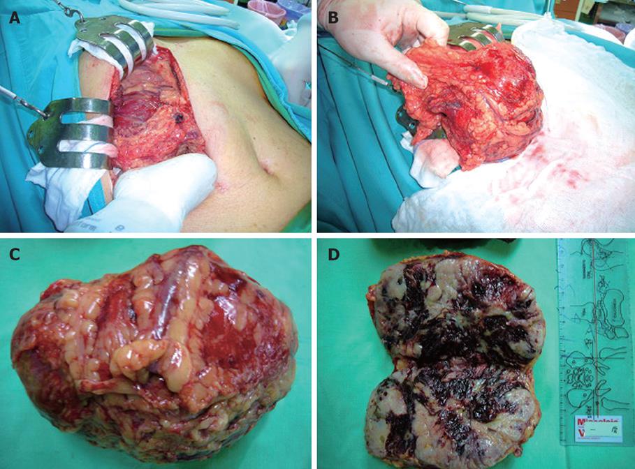
Figure 2 A huge tumor (12 cm × 8 cm × 6 cm) over the right upper quadrant area just below liver (A), blood supply of tumor from the omentum (B), and intraperitoneal tumor (C, D).
Figure 3 Two small mass lesions (3 cm × 2 cm and 2 cm × 1 cm) over S5 and S6, respectively.
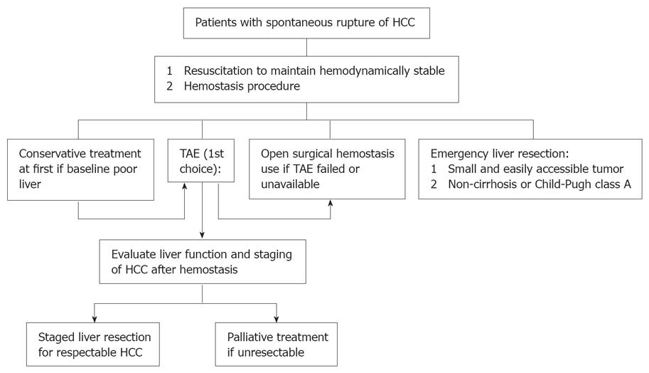
Figure 4 Logarithm about how to approach to the patient with spontaneously ruptured HCC.
- Citation: Hung MC, Wu HS, Lee YT, Hsu CH, Chou DA, Huang MH. Intraperitoneal metastasis of hepatocellular carcinoma after spontaneous rupture: A case report. World J Gastroenterol 2008; 14(24): 3927-3931
- URL: https://www.wjgnet.com/1007-9327/full/v14/i24/3927.htm
- DOI: https://dx.doi.org/10.3748/wjg.14.3927


