Copyright
©2008 The WJG Press and Baishideng.
World J Gastroenterol. Jun 28, 2008; 14(24): 3919-3921
Published online Jun 28, 2008. doi: 10.3748/wjg.14.3919
Published online Jun 28, 2008. doi: 10.3748/wjg.14.3919
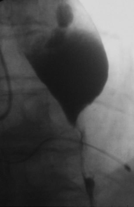
Figure 1 Radiographic image of the esophagus stenosis.
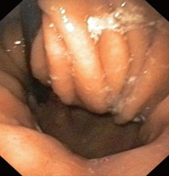
Figure 2 Endoscopic view of the hiatus hernia after passage through the tumor stenosis.
Figure 3 New stent design (Micro-Tech [Nanjing] Co.
Ltd., Nanjing, China; distributed by Leufen Medizintechnik, Aachen, Germany).
Figure 4 Radiographic image of the stent.
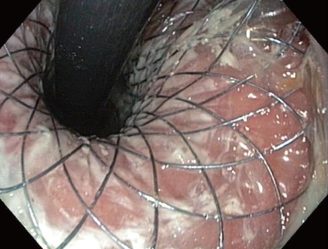
Figure 5 Endoscopic view of the distal end of the stent in inversion, 2 mo after treatment.
- Citation: Aymaz S, Dormann AJ. A new approach to endoscopic treatment of tumors of the esophagogastric junction with individually designed self-expanding metal stents. World J Gastroenterol 2008; 14(24): 3919-3921
- URL: https://www.wjgnet.com/1007-9327/full/v14/i24/3919.htm
- DOI: https://dx.doi.org/10.3748/wjg.14.3919


