Copyright
©2008 The WJG Press and Baishideng.
World J Gastroenterol. Jun 28, 2008; 14(24): 3903-3907
Published online Jun 28, 2008. doi: 10.3748/wjg.14.3903
Published online Jun 28, 2008. doi: 10.3748/wjg.14.3903
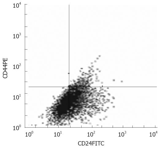
Figure 1 Analysis of Panc-1 pancreatic cancer cells by FACS.
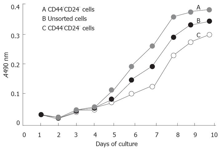
Figure 2 Growth curve of tumors cells in vitro.
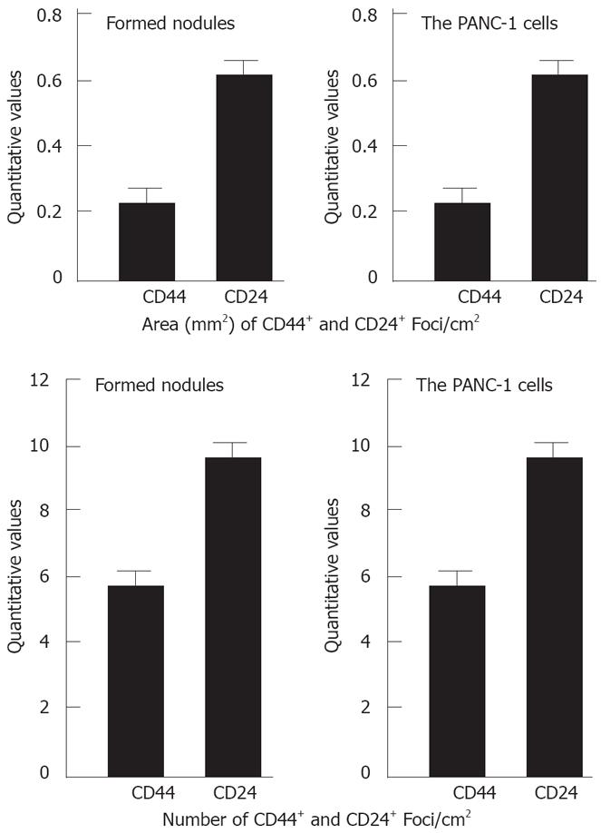
Figure 3 Quantitative values of CD44+ and CD24+ cell foci in the formed nodules and PANC-1 cells.
There was no significant difference between the formed nodules and PANC-1 cells.
- Citation: Huang P, Wang CY, Gou SM, Wu HS, Liu T, Xiong JX. Isolation and biological analysis of tumor stem cells from pancreatic adenocarcinoma. World J Gastroenterol 2008; 14(24): 3903-3907
- URL: https://www.wjgnet.com/1007-9327/full/v14/i24/3903.htm
- DOI: https://dx.doi.org/10.3748/wjg.14.3903


