Copyright
©2008 The WJG Press and Baishideng.
World J Gastroenterol. Jun 28, 2008; 14(24): 3812-3818
Published online Jun 28, 2008. doi: 10.3748/wjg.14.3812
Published online Jun 28, 2008. doi: 10.3748/wjg.14.3812
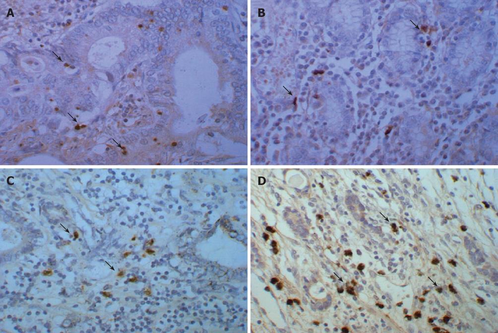
Figure 1 Expression and distribution of S100 protein and heparanase mRNA in gastric cancer tissues.
Immunohistochemical staining of S100 protein (× 400) with a high density of tumor infiltrating dendritic cells positively stained for S100 protein in the early stage gastric cancer tissues (A) and a low density of such cells in the late stage gastric cancer tissues (B), heparanase mRNA expression by in situ hybridization (× 400) with a low heparanase mRNA expression level in the early stage gastric cancer tissues (C) and a high heparanase mRNA expression level in the late stage gastric cancer tissues (D) (Arrows: Positively expressed cells).
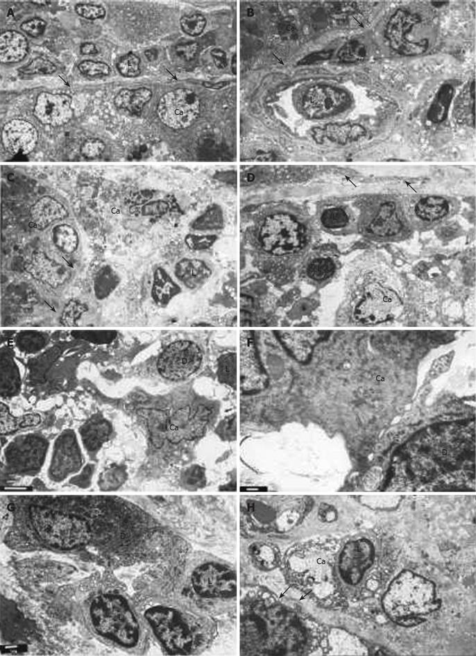
Figure 2 Transmission electron microscopy micrographs of the human gastric cancer tissues.
(A)-(D) showing the early stage cancer tissues (Arrows indicate basement membrane). A: The continuous basement membrane which consisted of the electron-dense outer layer and the electron-lucent inner layer was observed. The numerous tumor infiltrating lymphocytes (L) were located in one side of the basement membrane (× 2500); B and C: The intact basement membrane was found on the margin of cancer nests. The tumor infiltrating lymphocytes (L) appeared around the cancer cell (Ca) (B × 4000; C × 3000); D: The cancer cell (Ca) was surrounded by the tumor infiltrating lymphocytes (L), and the basement membrane is clearly visualized (× 2500); (E)-(H) showing the late stage cancer tissues. E: The relationships were displayed between cancer cells (Ca) or tumor infiltrating lymphocytes (L) and tumor infiltrating dendritic cells (D). Note the tumor with absent basement membrane (× 4000); F: A higher magnification of E exhibited the contact relationship between the cancer cell (Ca) and tumor infiltrating dendritic cell (D) (×20 000). G: The tumor infiltrating dendritic cell (D) was surrounded by several tumor infiltrating lymphocytes (L), and formed the dendritic cell-lymphocyte cluster (× 5000); H: The tumor infiltrating lymphocyte (L) appeared near the cancer cell (Ca), and the discontinuous or defective basement membrane of cancer nest can also be seen (double arrow) (× 5000).
- Citation: Xie ZJ, Liu Y, Jia LM, He YC. Heparanase expression, degradation of basement membrane and low degree of infiltration by immunocytes correlate with invasion and progression of human gastric cancer. World J Gastroenterol 2008; 14(24): 3812-3818
- URL: https://www.wjgnet.com/1007-9327/full/v14/i24/3812.htm
- DOI: https://dx.doi.org/10.3748/wjg.14.3812


