Copyright
©2008 The WJG Press and Baishideng.
World J Gastroenterol. Jun 21, 2008; 14(23): 3763-3767
Published online Jun 21, 2008. doi: 10.3748/wjg.14.3763
Published online Jun 21, 2008. doi: 10.3748/wjg.14.3763
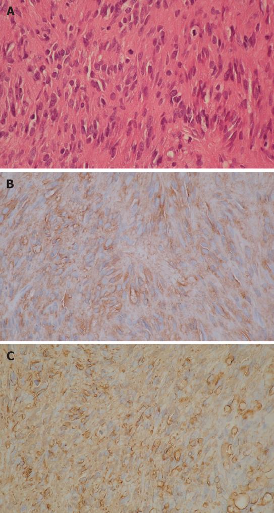
Figure 1 Histological study showing spindle cells with mitoses (HE, × 200) (A) and immunohistochemistry findings revealing positive staining for CD117 (B) and CD34 (× 200) (C) in primary GIST of the stomach.
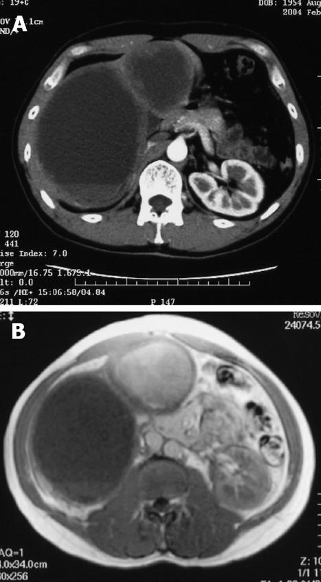
Figure 2 Contrast-enhanced CT scan (A) and MRI (B) on T1-weighted image 107 mo after the initial operation.
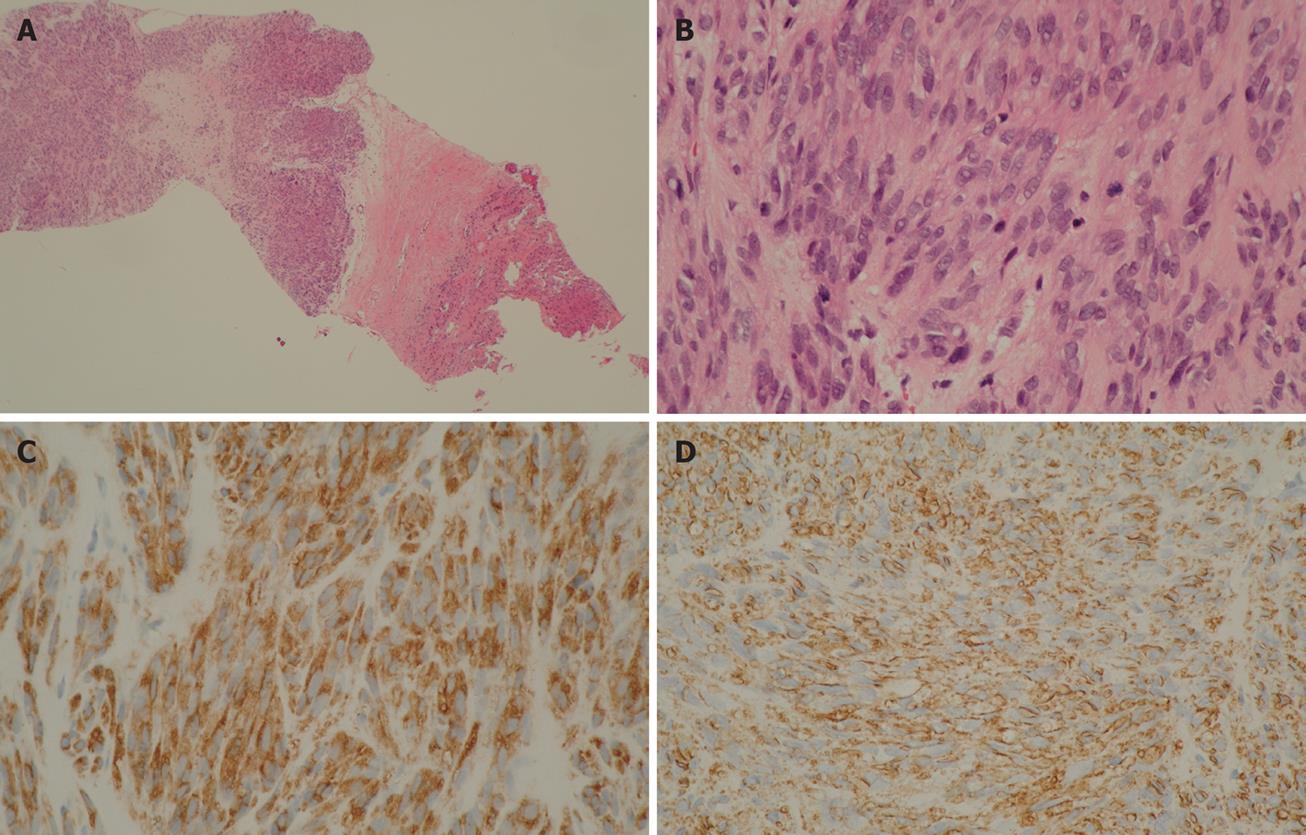
Figure 3 Scanning view of metastatic GIST (× 15) (A), histological study revealed spindle cells with mitoses (HE, × 200) (B), immunohistochemistry findings revealed positive staining for CD117 (C) and CD34 (× 200) (D) in metastatic GIST of the liver obtained from liver biopsy.
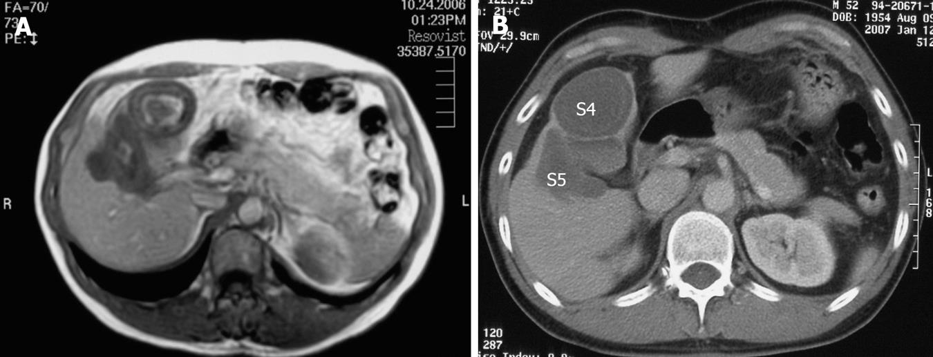
Figure 4 MRI on T1-WI 30 mo after treatment with Imatinib (A) and contrast-enhanced CT 34 mo after the treatment with Imatinib (B).
The metastatic lesions (S4 + S5) are indicated.
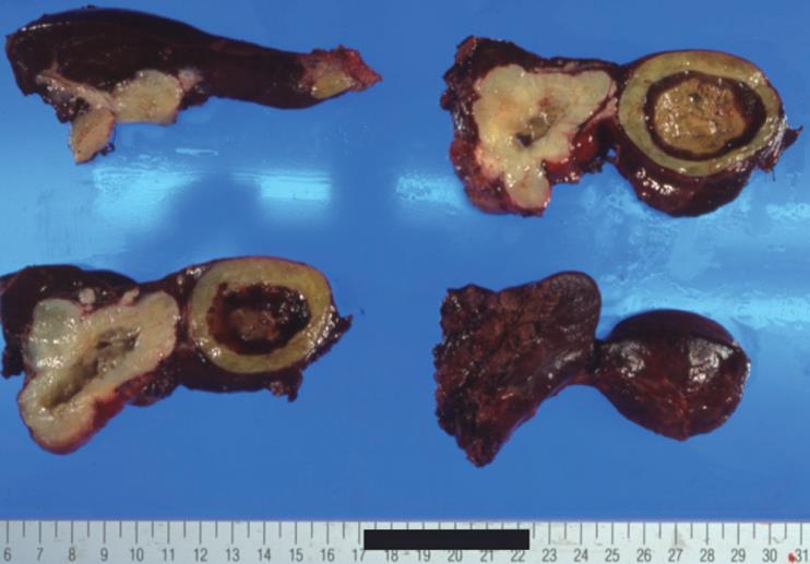
Figure 5 Serous and cut-surface views of resected specimen obtained from partial hepatectomy (S4 + S5) after treatment with Imatinib.
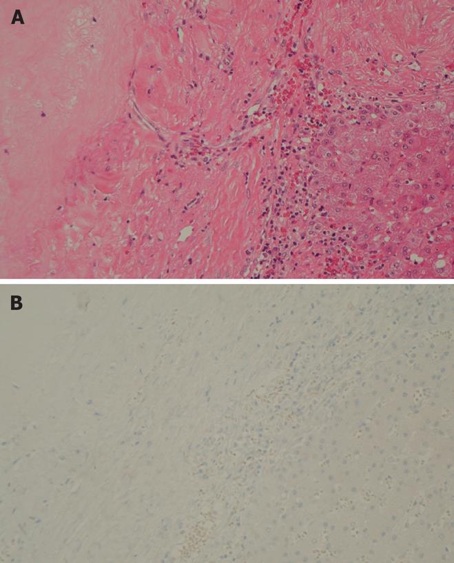
Figure 6 Histological study showing no viable tumor cells and hyaline degenerative tissues (HE, × 200) (A) and immunohistochemistry findings revealing negative staining for CD117 (× 200) (B) in the resected specimen after treatment with Imatinib.
- Citation: Suzuki S, Sasajima K, Miyamoto M, Watanabe H, Yokoyama T, Maruyama H, Matsutani T, Liu A, Hosone M, Maeda S, Tajiri T. Pathologic complete response confirmed by surgical resection for liver metastases of gastrointestinal stromal tumor after treatment with imatinib mesylate. World J Gastroenterol 2008; 14(23): 3763-3767
- URL: https://www.wjgnet.com/1007-9327/full/v14/i23/3763.htm
- DOI: https://dx.doi.org/10.3748/wjg.14.3763


