Copyright
©2008 The WJG Press and Baishideng.
World J Gastroenterol. Jun 14, 2008; 14(22): 3598-3601
Published online Jun 14, 2008. doi: 10.3748/wjg.14.3598
Published online Jun 14, 2008. doi: 10.3748/wjg.14.3598
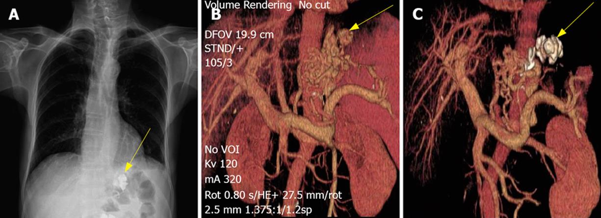
Figure 1 Subsequent chest X-ray on the 69-year-old male demonstrating marked retention of the cyanoacrylate/lipiodol mixture within the variceal bed in gastric fundus (arrow) (A), CTPA-reconstructed image showing abundant portal venous collaterals with no gastrorenal shunt and gastric fundal varices (arrow) communicating with the confluence through the left gastric vein (B) and shrinkage of gastric fundal varices and deposition of cyanoacrylate/lipiodol mixture (arrow), blocking the draining veins (C) after sclerotherapy.
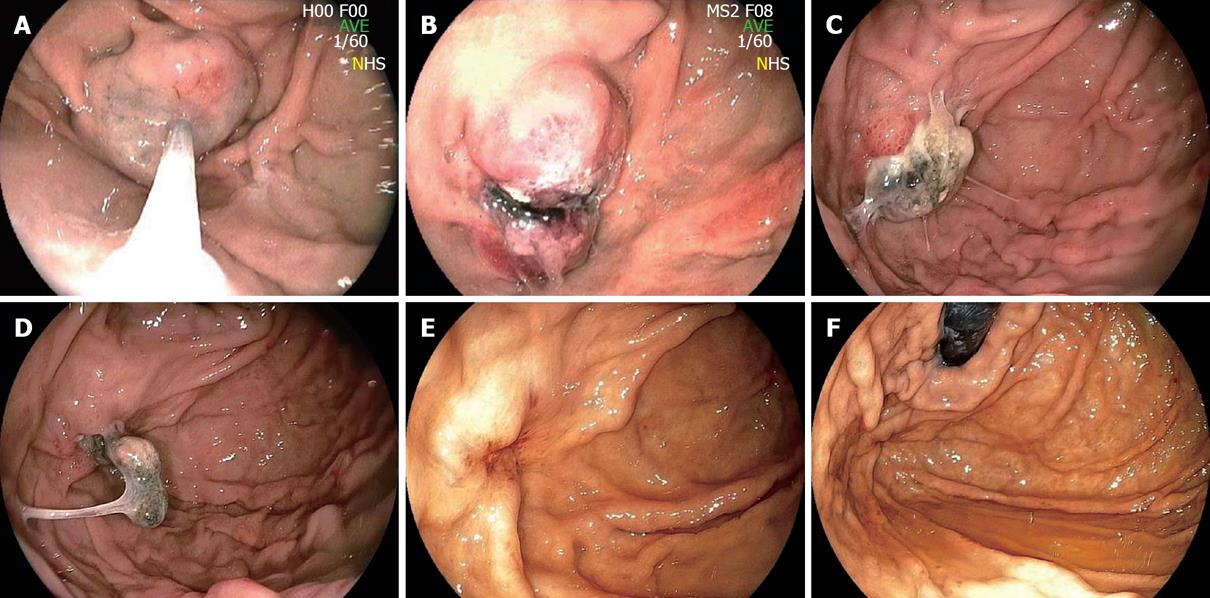
Figure 2 Endoscopy showing isolated gastric varices in gastric fundus (A) and abundant blood flow (B) in gastric varices before treatment, and shrinkage of gastric varices (C) and no blood flow signals (D), flat gastric varices with slight red spots on the top (E), and eradication of gastric varices (F) in the 69-year-old male patient after treatment.
Figure 3 Endoscopic findings after the first injection treatment with combined cyanoacrylate and aethoxysklerol (A), an marked ulcer noticed on the surface of varices 3 d after the first injection with no stigmata bleeding (B), an adhesive extrusion process 3 wk after the initial treatment (C), adhesive found on the surface of flat gastric varices 3 wk after the secondary injection (D), disappearance of the former massive gastric varices three and a half months after the secondary injection (E, F).
- Citation: Shi B, Wu W, Zhu H, Wu YL. Successful endoscopic sclerotherapy for bleeding gastric varices with combined cyanoacrylate and aethoxysklerol. World J Gastroenterol 2008; 14(22): 3598-3601
- URL: https://www.wjgnet.com/1007-9327/full/v14/i22/3598.htm
- DOI: https://dx.doi.org/10.3748/wjg.14.3598


