Copyright
©2008 The WJG Press and Baishideng.
World J Gastroenterol. Jun 14, 2008; 14(22): 3594-3597
Published online Jun 14, 2008. doi: 10.3748/wjg.14.3594
Published online Jun 14, 2008. doi: 10.3748/wjg.14.3594
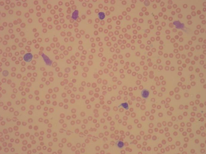
Figure 1 Peripheral blood film showing lymphocytosis and several smear cells.
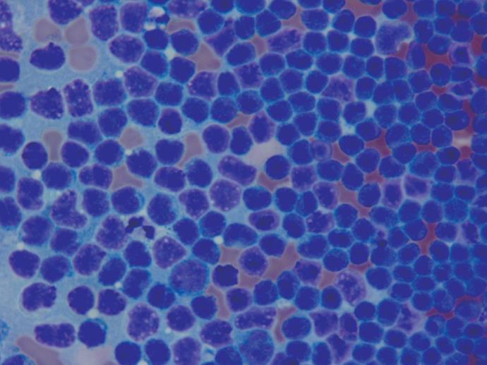
Figure 2 Cytospin of ascitic fluid showing infiltration by a large number of small, mature looking lymphocytes (DQ staining, × 400).
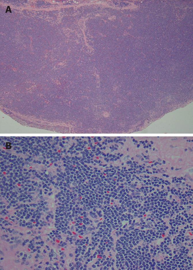
Figure 3 A: Lymph node section in low power showing total effacement of normal architecture (HE staining, × 40); B: Lymph node section in higher power showing infiltration with small lymphocytes (HE staining, × 400).
- Citation: Siddiqui N, Al-Amoudi S, Aleem A, Arafah M, Al-Gwaiz L. Massive ascites as a presenting manifestation of chronic lymphocytic leukemia. World J Gastroenterol 2008; 14(22): 3594-3597
- URL: https://www.wjgnet.com/1007-9327/full/v14/i22/3594.htm
- DOI: https://dx.doi.org/10.3748/wjg.14.3594


