Copyright
©2008 The WJG Press and Baishideng.
World J Gastroenterol. Jun 14, 2008; 14(22): 3587-3590
Published online Jun 14, 2008. doi: 10.3748/wjg.14.3587
Published online Jun 14, 2008. doi: 10.3748/wjg.14.3587
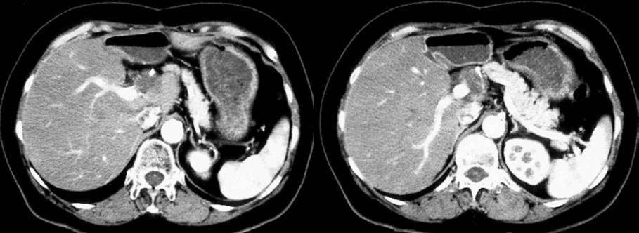
Figure 1 A CT during hepatic angiography showing a recurrent tumor with extensive invasion to the proper HA.
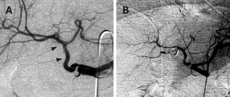
Figure 2 An angiogram of the celiac artery showing the proper and right arterial encasements (arrows) (A) and PRHA embolized with coils (B).
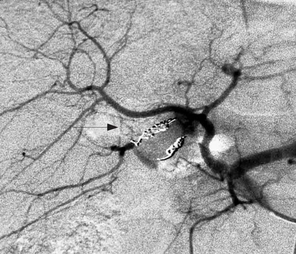
Figure 3 An embolized angiogram of the celiac artery 2 wk after enlargement of PRHA with its branches supplied from branches of the ARHA (arrow).
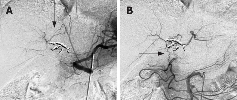
Figure 4 An angiogram of the celiac artery after the proper HA embolized with coils showing the collateral pathways from IPA to the branches of PRHA (arrow) (A), and an angiogram of the SMA after the proper hepatic artery embolized with coils showing the collateral pathways from SMA to the branches of ARHA (arrow) (B).
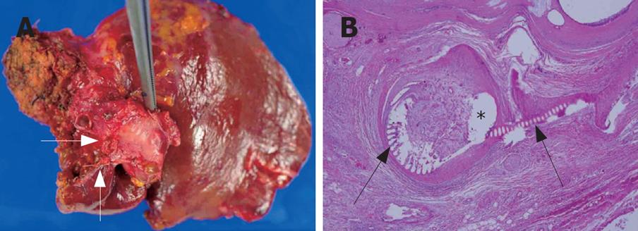
Figure 5 Tumor around the proper HA showing the anterior and posterior right hepatic arterial stump (arrows) (A) and histology revealing direct invasion of the tumor into the vascular wall of proper HA (*; HE staining, x 200) with traces of the coils in vascular lumen (arrow) (B).
- Citation: Miura T, Hakamada K, Ohata T, Narumi S, Toyoki Y, Nara M, Ishido K, Ohashi M, Akasaka H, Jin H, Kubo N, Ono S, Kijima H, Sasaki M. Resection of a locally advanced hilar tumor and the hepatic artery after stepwise hepatic arterial embolization: A case report. World J Gastroenterol 2008; 14(22): 3587-3590
- URL: https://www.wjgnet.com/1007-9327/full/v14/i22/3587.htm
- DOI: https://dx.doi.org/10.3748/wjg.14.3587


