Copyright
©2008 The WJG Press and Baishideng.
World J Gastroenterol. Jun 14, 2008; 14(22): 3563-3568
Published online Jun 14, 2008. doi: 10.3748/wjg.14.3563
Published online Jun 14, 2008. doi: 10.3748/wjg.14.3563
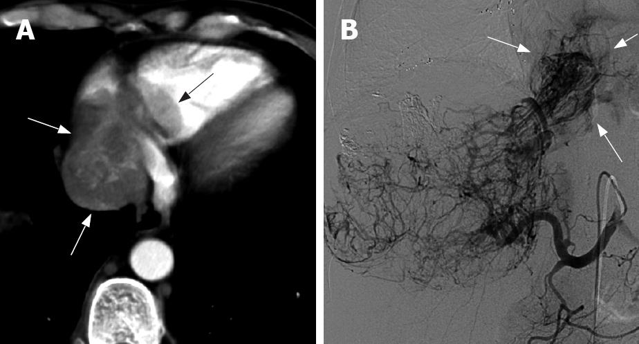
Figure 1 A 52-year-old man with a big massive HCC located in the right lobe, the tumor encroaches the PV and IVC and intrudes into the RA.
The embolus increased 5 cm within 3 mo. A: Arterial phase of CT scan shows a well-defined lobulated filling defect (6.7 cm x 7.5 cm) in the RA (arrow) and an irregular “stick”-like enhancement; B: Angiographic image in the second time TACE shows that the tumor is a hypervascular lesion and the artery enters the RA by passing the IVC, and is a “grating”-like type (arrow).
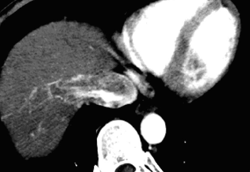
Figure 2 A 42-year-old man with a massive HCC located in the right lobe, the tumor encroaches the HV and IVC and intrudes into the RA.
Arterial phase of CT scan shows a “stick”-like enhanced arterial vessel entering the entrance of the RA.
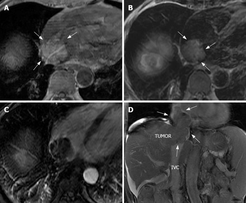
Figure 3 A 69-year-old man with a multinodular HCC located in the right lobe, 7 mo after the 6th TACE, embolus in the RA increased to 4 cm (A-C are the same patient).
A: T1-weighted image shows a higher signal nodular (TR: 195 ms/TE: 4.2 ms, ST: 8.0 mm) in RA (arrow); B: T2-weighted image shows a higher signal nodular of HCC in the right diaphragmatic dome, the same well-defined higher signal nodular in the RA and a low signal around the core-cavity (TR: 7058.8 ms/TE: 89.2 ms, ST: 8.0 mm); C: The arterial phase image shows an embolus with low signal filling defect and a “stick”-like enhancement (TR: 190 ms/TE: 1.9 ms, ST: 8.0 mm); D: The coronal image shows the tumor entering into the RA via the widened IVC, the IVC lumen was almost completely filled (arrow) (TR: 3.7 ms/TE: 1.6 ms, ST: 7.0 mm).
Figure 4 A 49-year-old man with multinodular recurrence after HCC operation, with the embolus of RA discovered at the same time.
The patient underwent TACE twice. A: The portal phase T1-weighted image shows a low signal embolus entering the IVC from middle HV (arrow) (TR: 220 ms/TE: 18ms, ST: 8.0 mm); B: The coronal image shows a well-defined “bottle-gourd” -shape filling defect in RA (arrow) (TR: 3.3 ms/TE: 1.5 ms/TI: 7.0 ms, ST: 3.0 mm); C: The image after perfused Lipiodol shows a nodular and lump tumor of the right and left lobe filled with Lipiodol, which was also retained in the embolus of RA (arrow).
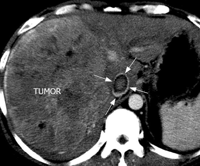
Figure 5 A 46-year-old man with a large mass of HCC in the right lobe.
The intra-liver tumor entered the RA by IVC. Cross sectional CT scan in portal phase shows a well-defined filling defect in IVC and a “ring”-like sign (arrow).
- Citation: Cheng HY, Wang XY, Zhao GL, Chen D. Imaging findings and transcatheter arterial chemoembolization of hepatic malignancy with right atrial embolus in 46 patients. World J Gastroenterol 2008; 14(22): 3563-3568
- URL: https://www.wjgnet.com/1007-9327/full/v14/i22/3563.htm
- DOI: https://dx.doi.org/10.3748/wjg.14.3563


