Copyright
©2008 The WJG Press and Baishideng.
World J Gastroenterol. Jun 14, 2008; 14(22): 3490-3496
Published online Jun 14, 2008. doi: 10.3748/wjg.14.3490
Published online Jun 14, 2008. doi: 10.3748/wjg.14.3490
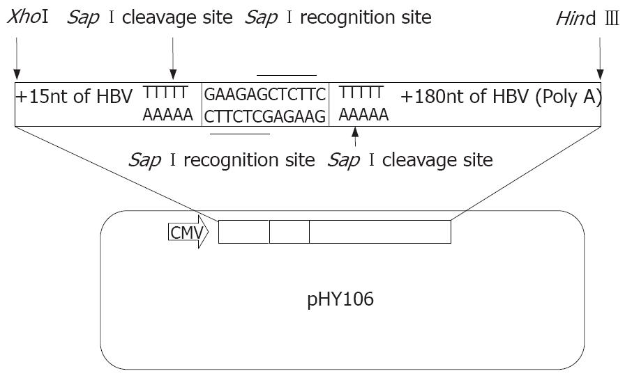
Figure 1 The schematic map of HBV expression vector of pHY106.
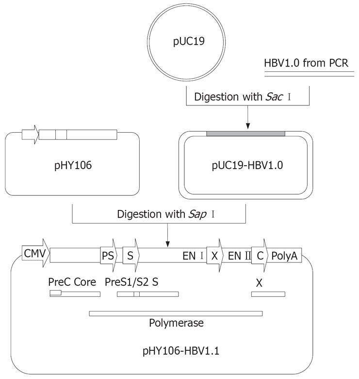
Figure 2 Construction of replicons of clinical HBV isolates using pHY106.
Full-length HBV genomes from PCR amplication can be digested with Sap I and inserted into Sap I-digested pHY106 to produce HBV replicon. HBV open reading frames (ORFs) for the pre-core (PC), core, preS1 (PS1), preS2 (2) surface (S), polymerase, and X genes are indicated by square frame. Promoter sequences for the preS (PS), surface (S) and core (C) genes are indicated by arrows.
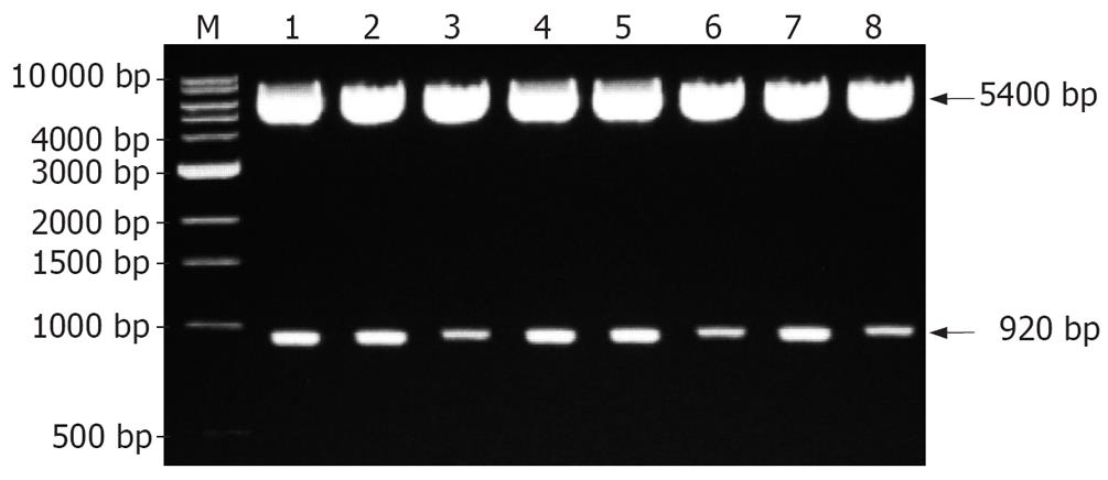
Figure 3 Identification of recombinant expression plasmids by restriction endonuclease digestion analysis.
M: 1 kb DNA ladder marker; Lane 1-8: Recombinant plasmids digested with Hind III and Nsi I.
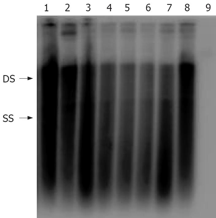
Figure 4 Analysis of intracellular replication of clinical HBV isolates.
HBV isolates were cloned into the vector pHY106 and transfected into Huh2 cells. Seventy-two hours after transfection, intracellular replicative intermediates were isolated and analyzed by Southern blot. Double-stranded (DS) and single-stranded (SS) intermediates are indicated. Lane 1-8: eight replicons of clinical HBV isolates; Lane 9: pHY106 negative control.
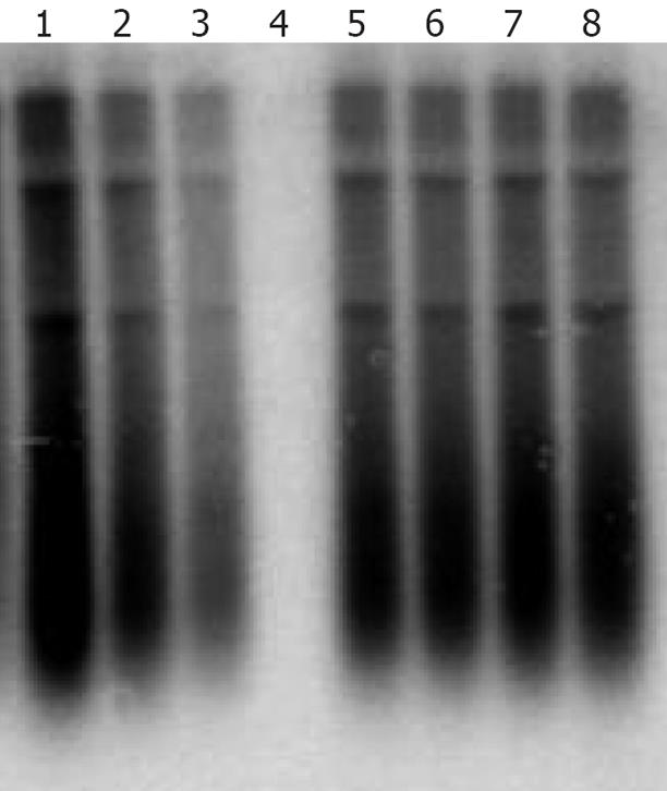
Figure 5 Antiviral susceptibility of an HBV isolate cloned from a patient with lamivudine resistance.
An HBV isolate amplified from the sera of patients failing in lamivudine therapy was cloned into the pHY106 vector and analyzed in vitro. Lane 1-4: transfected cells treated with 0.01, 0.1, 1 and 10 &mgr;mol/L of adefovir; Lane 5-8: transfected cells treated with 0.01, 0.1, 1 and 10 &mgr;mol/L of lamivudine.
- Citation: Lu YP, Guo T, Wang BJ, Dong JH, Zhu JF, Liu Z, Lu MJ, Yang DL. Replication of clinical hepatitis B virus isolate and its application for selecting antiviral agents for chronic hepatitis B patients. World J Gastroenterol 2008; 14(22): 3490-3496
- URL: https://www.wjgnet.com/1007-9327/full/v14/i22/3490.htm
- DOI: https://dx.doi.org/10.3748/wjg.14.3490


