Copyright
©2008 The WJG Press and Baishideng.
World J Gastroenterol. May 28, 2008; 14(20): 3269-3272
Published online May 28, 2008. doi: 10.3748/wjg.14.3269
Published online May 28, 2008. doi: 10.3748/wjg.14.3269
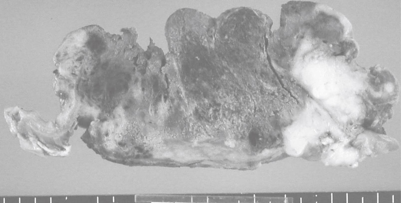
Figure 1 Cut surfaces of the tumor demonstrate two different features, a hemorrhagic brown area and a whitish-yellow area.
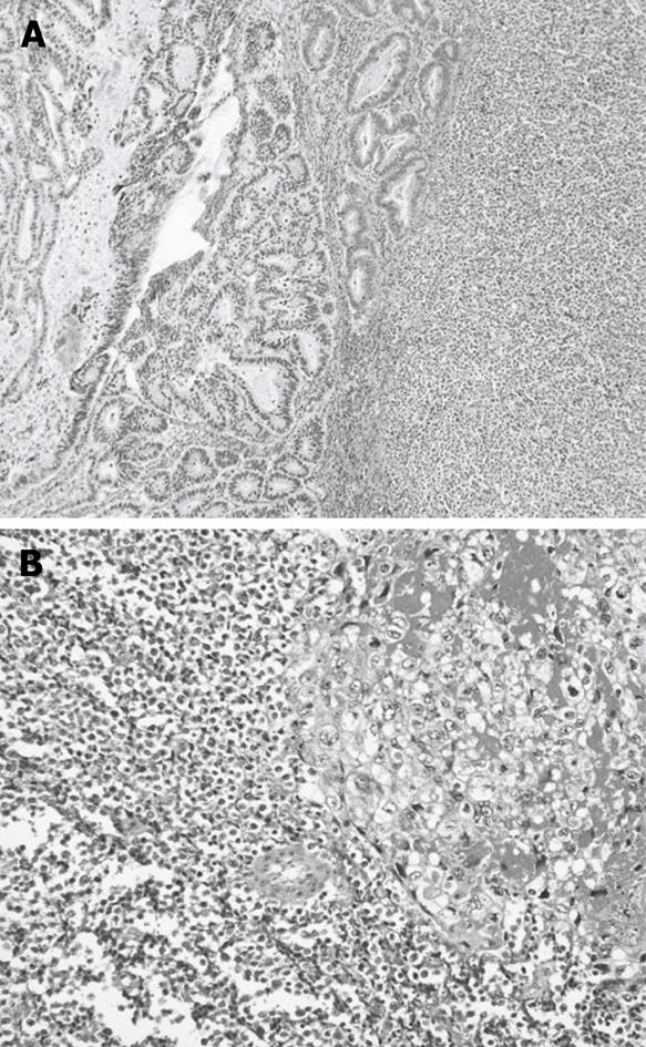
Figure 2 A: The hemorrhagic brown area is composed of choriocarcinoma; B: The whitish-yellow area contains small cell carcinoma.
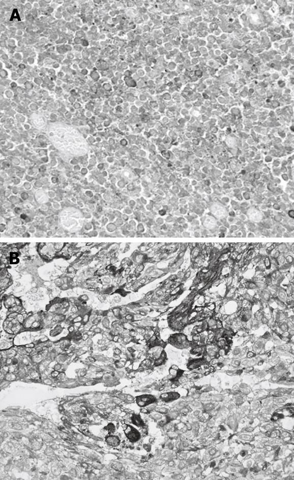
Figure 3 A: The choriocarcinomatous foci contain cells positive for B-hCG; B: The small cell carcinomatous foci contain cells positive for chromogranin A.
- Citation: Hirano Y, Hara T, Nozawa H, Oyama K, Ohta N, Omura K, Watanabe G, Niwa H. Combined choriocarcinoma, neuroendocrine cell carcinoma and tubular adenocarcinoma in the stomach. World J Gastroenterol 2008; 14(20): 3269-3272
- URL: https://www.wjgnet.com/1007-9327/full/v14/i20/3269.htm
- DOI: https://dx.doi.org/10.3748/wjg.14.3269


