Copyright
©2008 The WJG Press and Baishideng.
World J Gastroenterol. May 28, 2008; 14(20): 3266-3268
Published online May 28, 2008. doi: 10.3748/wjg.14.3266
Published online May 28, 2008. doi: 10.3748/wjg.14.3266
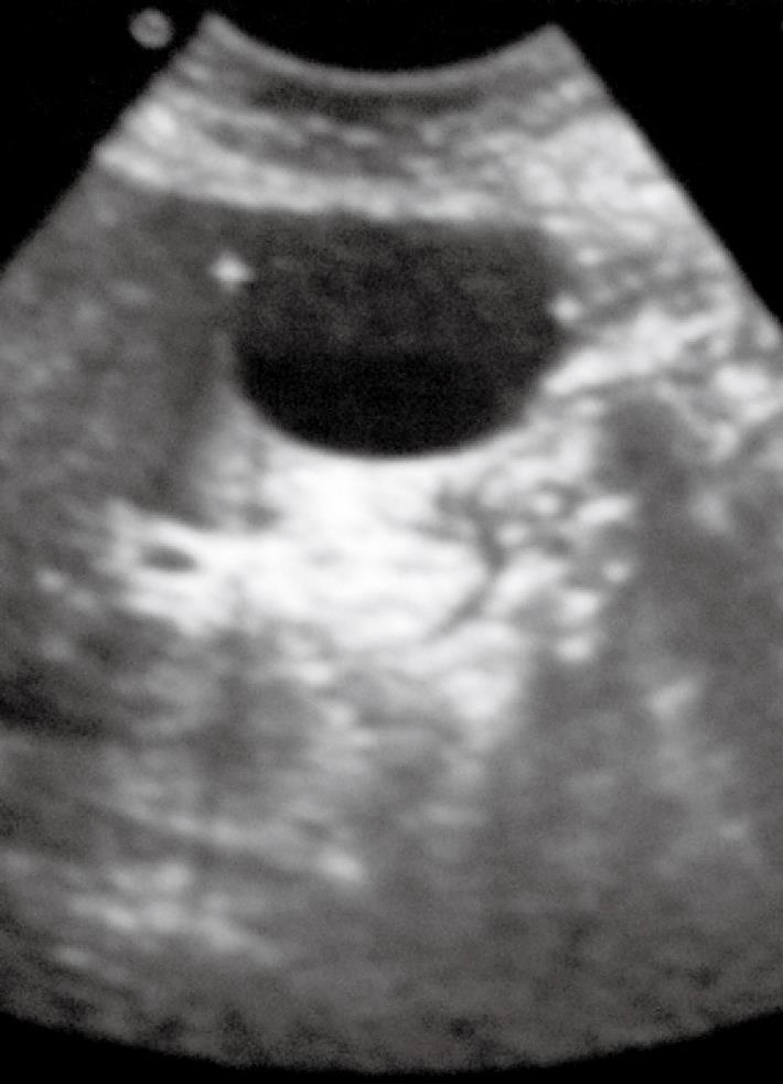
Figure 1 Ultrasound appearance of the cystic abdominal mass.
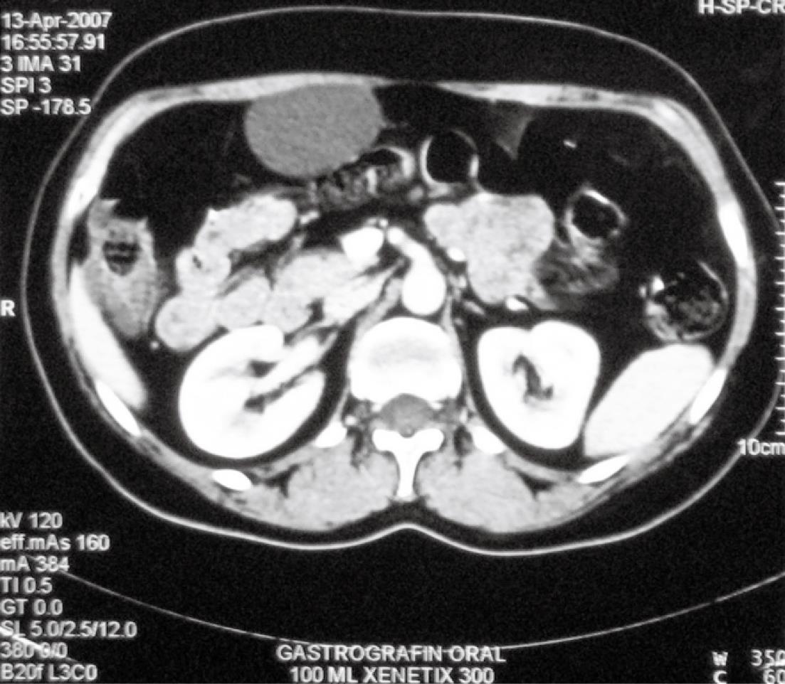
Figure 2 Abdominal CT scan with intravenous contrast media used showing a water-density mass attached to the anterior abdominal wall.
A well circumscribed area of low attenuation in contact with the abdominal wall is identified.
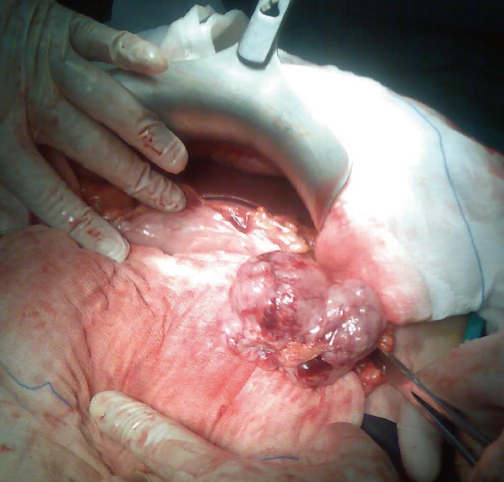
Figure 3 The cyst is shown originating from the ligamentum teres.
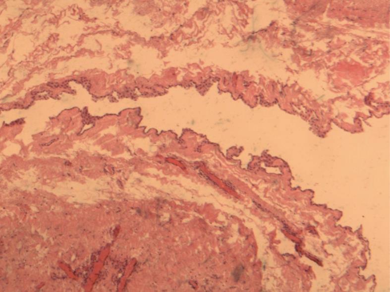
Figure 4 Hematoxylin-eosin staining of the cyst wall shows the single layered cuboidal epithelium (× 40).
- Citation: Lagoudianakis EE, Michalopoulos N, Markogiannakis H, Papadima A, Filis K, Kekis P, Katergiannakis V, Manouras A. A symptomatic cyst of the ligamentum teres of the liver: A case report. World J Gastroenterol 2008; 14(20): 3266-3268
- URL: https://www.wjgnet.com/1007-9327/full/v14/i20/3266.htm
- DOI: https://dx.doi.org/10.3748/wjg.14.3266


