Copyright
©2008The WJG Press and Baishideng.
World J Gastroenterol. Jan 14, 2008; 14(2): 211-217
Published online Jan 14, 2008. doi: 10.3748/wjg.14.211
Published online Jan 14, 2008. doi: 10.3748/wjg.14.211
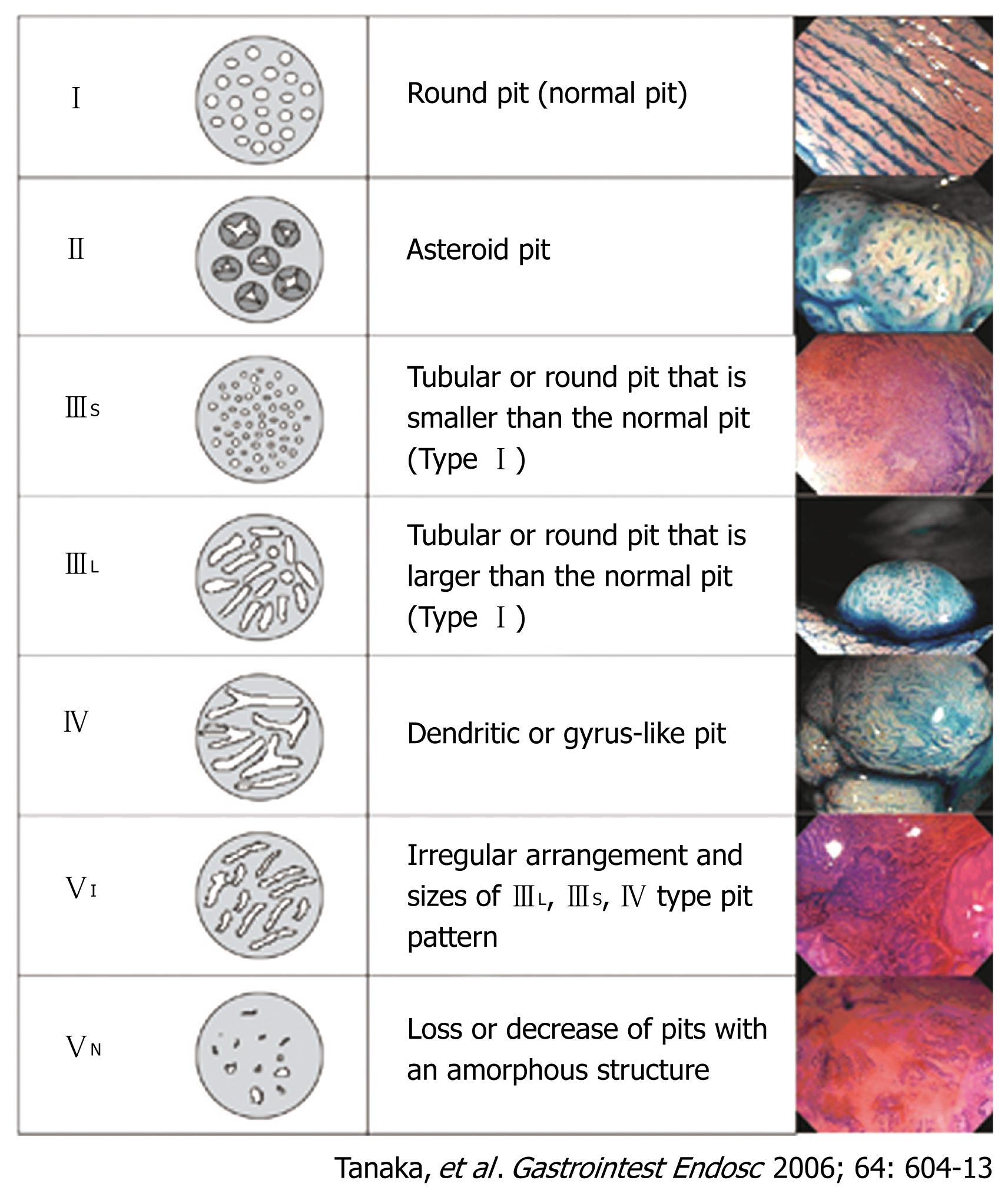
Figure 1 Classification of pit patterns of colorectal lesions.
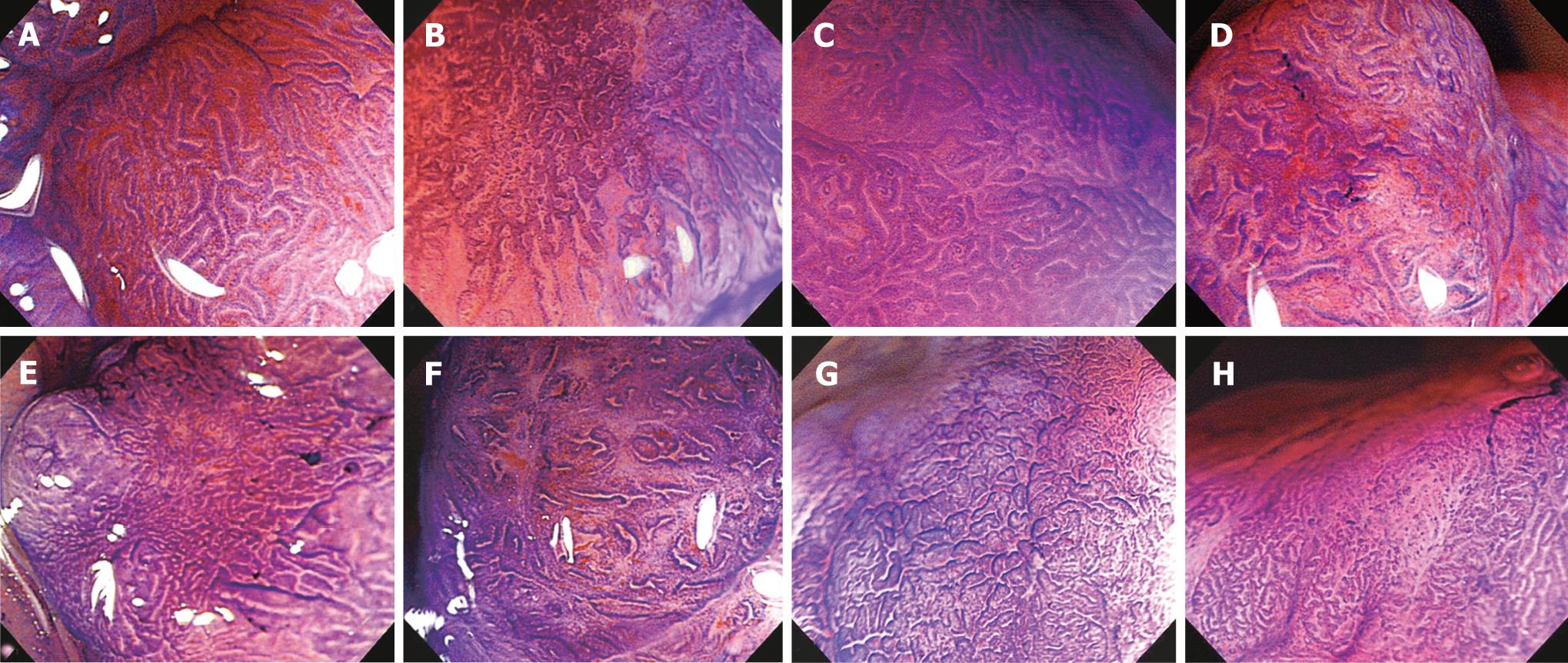
Figure 2 Magnifying features of colorectal neoplasm (crystal violet): A: Regular pit margins; B: Irregular pit margins; C: Clear staining characteristics of the areas between pits; D: Unclear staining characteristics of the areas between pits; E: High residual pit density; F: Low residual pit density; G: Narrow intervening membrane between pits; H: Wide intervening membrane between pits.
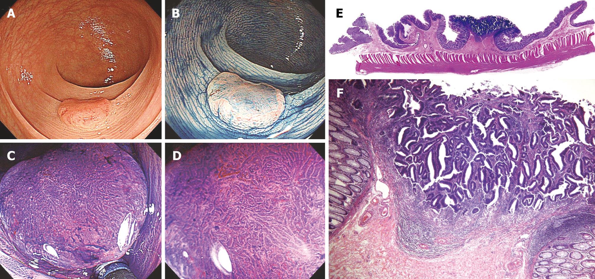
Figure 3 Type IIa + 10IIc lesion, 12 mm in diameter.
A: Standard colonoscopic view; B: Standard colonoscopic view with indigo carmine spraying; C, D: Magnifying colonoscopic picture with crystal violet staining reveals type VI pit pattern. Irregular pit margins, unclear staining characteristics of the areas between pits, > 5 mm area of type VI pit pattern, high residual pit density, and narrow intervening membrane between pits is revealed; E: Cross-section (hematoxylin-eosin, × 8) of a surgically resected specimen showing submucosal invasion (1800 &mgr;m); F: Low-power view (hematoxylin-eosin, × 40) of type VI pit pattern. Muscularis mucosae have disappeared. Desmoplastic reactions are mild to moderate. In this case, lymph node metastasis was not detected.
- Citation: Kanao H, Tanaka S, Oka S, Kaneko I, Yoshida S, Arihiro K, Yoshihara M, Chayama K. Clinical significance of type VI pit pattern subclassification in determining the depth of invasion of colorectal neoplasms. World J Gastroenterol 2008; 14(2): 211-217
- URL: https://www.wjgnet.com/1007-9327/full/v14/i2/211.htm
- DOI: https://dx.doi.org/10.3748/wjg.14.211


