Copyright
©2008 The WJG Press and Baishideng.
World J Gastroenterol. May 21, 2008; 14(19): 3098-3100
Published online May 21, 2008. doi: 10.3748/wjg.14.3098
Published online May 21, 2008. doi: 10.3748/wjg.14.3098
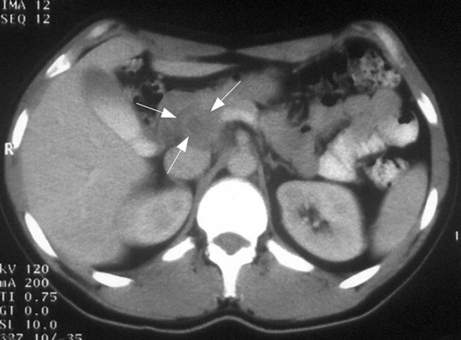
Figure 1 Abdominal CT-scan showing a low density mass on the posterior aspect of the head of the pancreas with contrast enhancing solid rim (arrow).
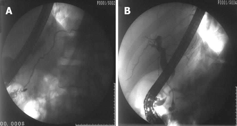
Figure 2 A: ERCP with a normal pancreatogram; B: A smooth long severe narrowing of the distal common bile duct.
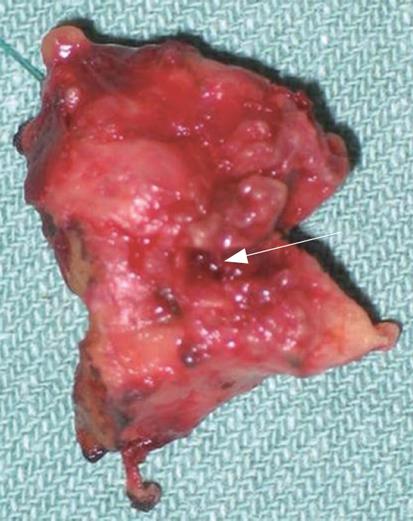
Figure 3 The resected part of the common bile duct with fistula on the posterior wall (arrow).
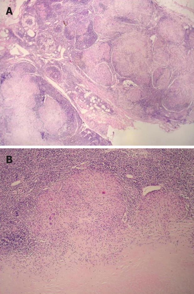
Figure 4 A: Extensive chronic granulomatous lymphadenitis (HE, × 13); B: Focal tuberculoid granuloma formation (HE, × 64).
- Citation: Colovic R, Grubor N, Jesic R, Micev M, Jovanovic T, Colovic N, Atkinson HD. Tuberculous lymphadenitis as a cause of obstructive jaundice: A case report and literature review. World J Gastroenterol 2008; 14(19): 3098-3100
- URL: https://www.wjgnet.com/1007-9327/full/v14/i19/3098.htm
- DOI: https://dx.doi.org/10.3748/wjg.14.3098


