Copyright
©2008 The WJG Press and Baishideng.
World J Gastroenterol. May 21, 2008; 14(19): 3095-3097
Published online May 21, 2008. doi: 10.3748/wjg.14.3095
Published online May 21, 2008. doi: 10.3748/wjg.14.3095
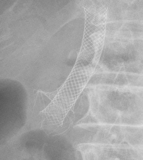
Figure 1 Simple abdomen examination demonstrating compression of deformed distal tip of the outer biliary metallic stent shooting out radially in all directions.
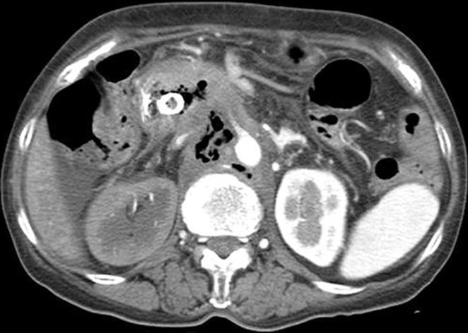
Figure 2 Abdominal CT scan showing biliary metallic stents with a lesion arising from the pancreatic head and the trajectory of air bubble densities traced from the pancreatic head to the lower paraaortic lesions.
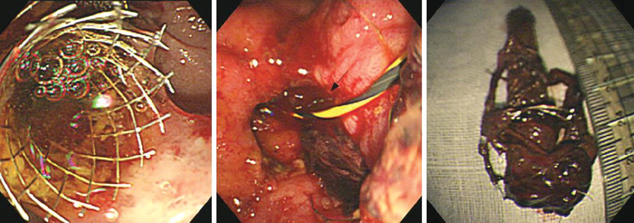
Figure 3 EGD.
A: On previous admission (one year ago), placement of a stent into a stent due to clogging; B: On the present admission, a circular hole with bleeding (arrow) caused by stent-induced perforation following removal of stents; C: Retrieved biliary metallic stents showing deformed barbs of the uncovered wall stent tip on the distal portion of the covered wall stent.
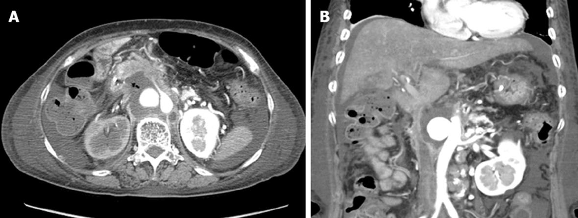
Figure 4 Abdominal CT scan showing decreased air densities in the pancreatic head to the paraaortic area (A) and a circular contrast collecting aneurysm of aorta (B).
- Citation: Lee TH, Park DH, Park JY, Lee SH, Chung IK, Kim HS, Park SH, Kim SJ. Aortoduodenal fistula and aortic aneurysm secondary to biliary stent-induced retroperitoneal perforation. World J Gastroenterol 2008; 14(19): 3095-3097
- URL: https://www.wjgnet.com/1007-9327/full/v14/i19/3095.htm
- DOI: https://dx.doi.org/10.3748/wjg.14.3095


