Copyright
©2008 The WJG Press and Baishideng.
World J Gastroenterol. May 21, 2008; 14(19): 3092-3094
Published online May 21, 2008. doi: 10.3748/wjg.14.3092
Published online May 21, 2008. doi: 10.3748/wjg.14.3092
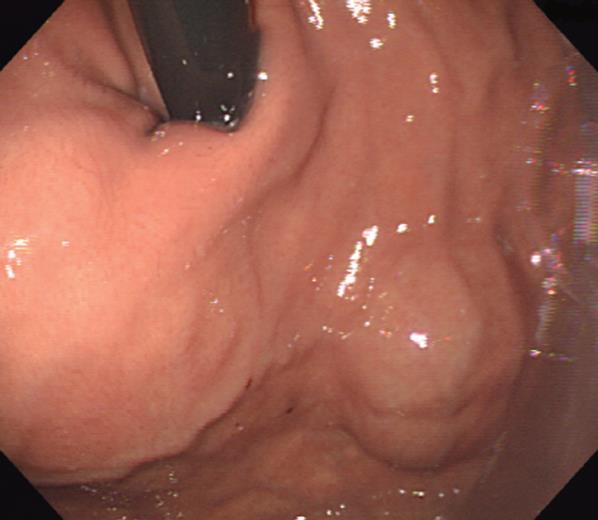
Figure 1 Endoscopic photograph demonstrating a protruding mass on the cardia of stomach.
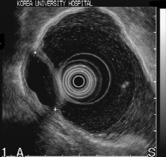
Figure 2 EUS showing a hypoechoic mass (3.
6 cm in diameter) which was suspicious of a gastric duplication cyst or a GIST.
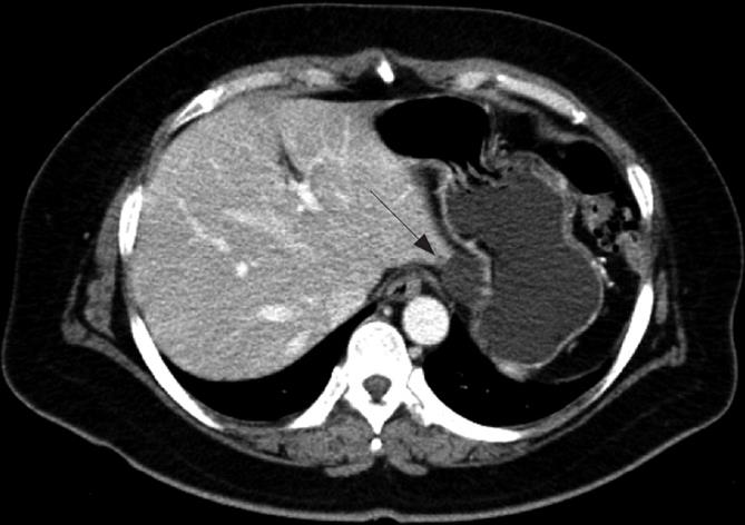
Figure 3 Abdominal CT scan showing a low density lesion in the submucosal layer of the gastric cardia (arrow).
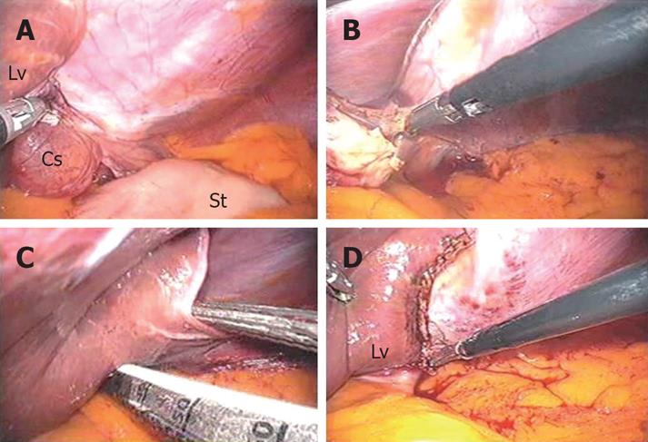
Figure 4 Operative procedures for the hepatic cyst in the left lateral segment of liver (A), after dissection of the triangular ligament (B), liver wedge resection using an endoscopic linear stapler (C, D).
Lv: Liver, Cs: Hepatic cyst, St: Stomach.
- Citation: Park JM, Kim J, Kim HI, Kim CS. Hepatic cyst misdiagnosed as a gastric submucosal tumor: A case report. World J Gastroenterol 2008; 14(19): 3092-3094
- URL: https://www.wjgnet.com/1007-9327/full/v14/i19/3092.htm
- DOI: https://dx.doi.org/10.3748/wjg.14.3092


