Copyright
©2008 The WJG Press and Baishideng.
World J Gastroenterol. May 21, 2008; 14(19): 3074-3080
Published online May 21, 2008. doi: 10.3748/wjg.14.3074
Published online May 21, 2008. doi: 10.3748/wjg.14.3074
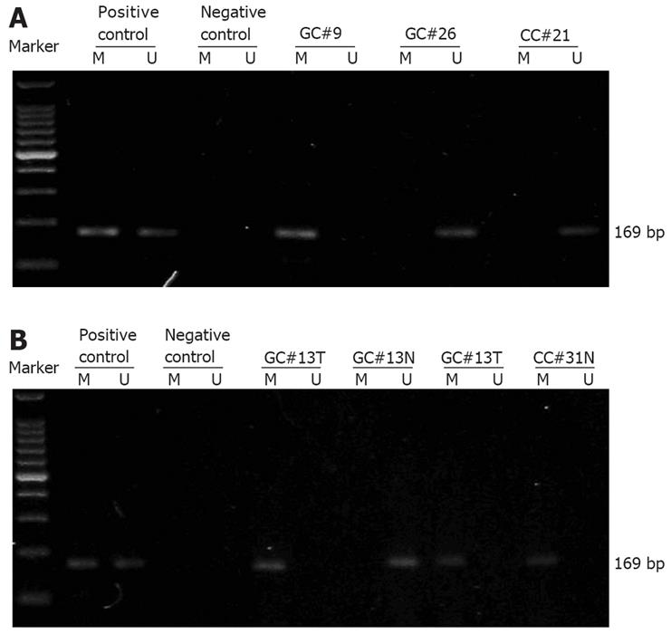
Figure 1 Representative results showing RASSF1A promoter methylation status identified by MSPCR in gastric and adenocarcinoma patients.
Identification of RASSF1A promoter methylation status in serum samples from gastric and colorectal adenocarcinoma patients (A) and in paired tumor and adjacent normal tissue from gastric and colorectal adenocarcinoma patients (B). A 100-bp DNA ladder marker (TaKaRa, Shiga, Japan) was used. Lanes M and U indicate the amplified products with primers recognizing specific methylated and unmethylated sequences, respectively. GC: Gastric adenocarcinoma; CC: Colorectal adenocarcinoma; T: Tumor tissue; N: Paired adjacent normal tissue.
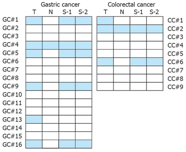
Figure 2 Comparison of RASSF1A promoter methylation status in tissue and serum samples.
For each patient, the RASSF1A promoter methylation status was analyzed in tumor tissue (T), adjacent normal tissue (N), preoperative serum (S-1), and postoperative serum collected 4 wk after surgery (S-2). Solid boxes indicate methylation, blank ones indicate unmethylation of RASSF1A promoter. GC: Gastric adenocarcinoma; CC: Colorectal adenocarcinoma.
-
Citation: Wang YC, Yu ZH, Liu C, Xu LZ, Yu W, Lu J, Zhu RM, Li GL, Xia XY, Wei XW, Ji HZ, Lu H, Gao Y, Gao WM, Chen LB. Detection of
RASSF1A promoter hypermethylation in serum from gastric and colorectal adenocarcinoma patients. World J Gastroenterol 2008; 14(19): 3074-3080 - URL: https://www.wjgnet.com/1007-9327/full/v14/i19/3074.htm
- DOI: https://dx.doi.org/10.3748/wjg.14.3074


