Copyright
©2008 The WJG Press and Baishideng.
World J Gastroenterol. May 21, 2008; 14(19): 2995-2999
Published online May 21, 2008. doi: 10.3748/wjg.14.2995
Published online May 21, 2008. doi: 10.3748/wjg.14.2995
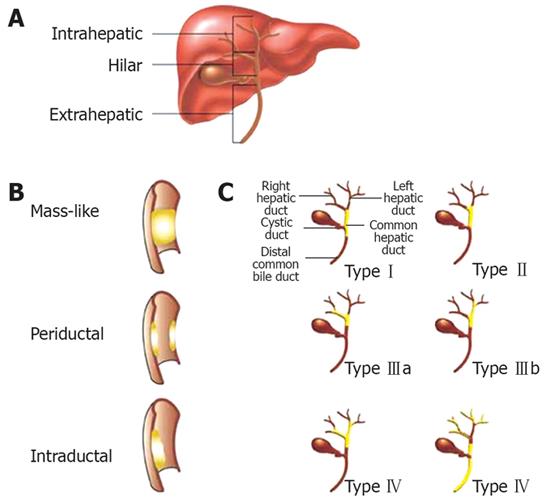
Figure 1 Classification of cholangiocarcinoma.
A: The classification of cholangiocarcinoma can be based on anatomic location, intrahepatic, hilar or extrahepatic; B: Nonhilar lesions can be described as mass-like, periductal or intraductal; C: Bismuth classification for hilar lesions.
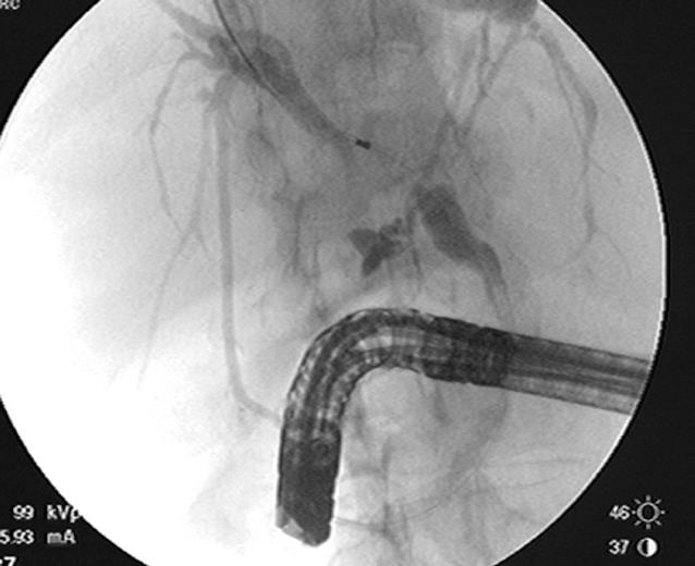
Figure 2 Hilar lesion causing bilateral strictures at the bifurcation of the left and right hepatic ducts with proximal dilation.
Figure 3 A: Distal common bile duct lesion with proximal biliary dilation; B: Fine needle aspiration of common bile duct lesion; C: ERCP shows distal common bile duct stricture consistent with findings on EUS.
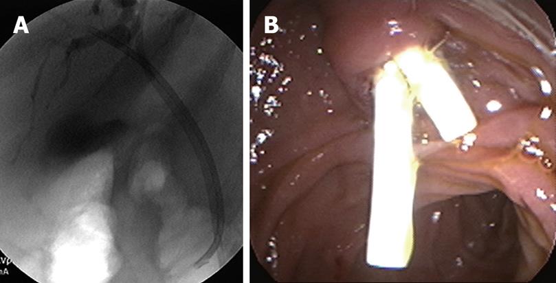
Figure 4 A: In this patient with a hilar mass, double stents were placed within the right and left hepatic systems to allow adequate drainage of contrast after cholangiogram was performed to decrease the risk of cholangitis; B: Endoscopic view of bilateral stents placed.
- Citation: Nguyen K, Jr JTS. Review of endoscopic techniques in the diagnosis and management of cholangiocarcinoma. World J Gastroenterol 2008; 14(19): 2995-2999
- URL: https://www.wjgnet.com/1007-9327/full/v14/i19/2995.htm
- DOI: https://dx.doi.org/10.3748/wjg.14.2995


