Copyright
©2008 The WJG Press and Baishideng.
World J Gastroenterol. May 14, 2008; 14(18): 2924-2927
Published online May 14, 2008. doi: 10.3748/wjg.14.2924
Published online May 14, 2008. doi: 10.3748/wjg.14.2924
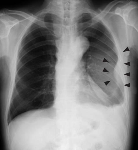
Figure 1 Chest radiography revealed massive plural effusion and a tumor elevating smoothly from the left chest wall (arrowheads), near which the fifth rib could not be traced.
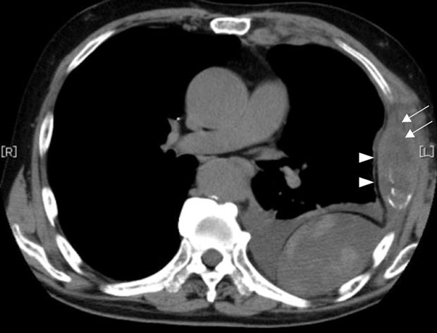
Figure 2 Chest CCT depicted a mass with necrosis inside the tumor (arrows) and enhancement of its border (arrowheads), and extrapleural tumor and pleural effusion.
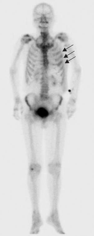
Figure 3 Bone scintigraphy showed an abnormal opaque uptake in the left third to fifth ribs (arrows).
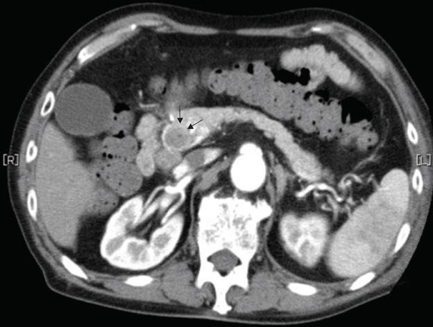
Figure 4 Abdominal CT revealed a gradually enhanced tumor in the distal bile duct (arrows), without any regional lymph node or liver metastasis.
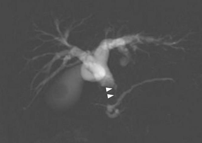
Figure 5 MRCP revealed stenosis (arrow heads) of the distal common bile duct.
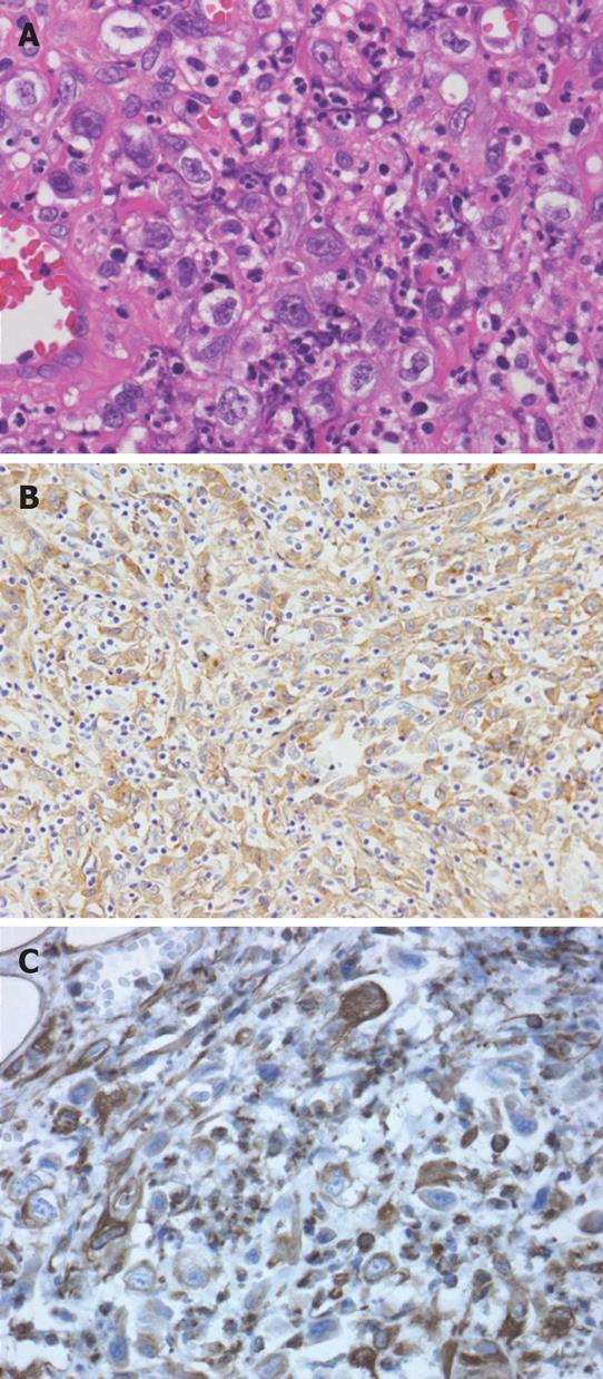
Figure 6 Histological examination of the resected chest wall showed scattered atypical cells among numerous inflammatory cells (A).
Immunohistochemistry for pan-cytokeratin as an epithelial cell marker (B) and vimentin as a mesenchymal cell marker (C) both showed positivity in these atypical cells.
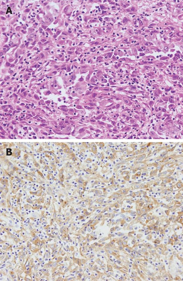
Figure 7 Histological examination revealed a poorly differentiated adenocarcinoma with sarcomatous change in the distal bile duct, which slightly invaded into the pancreas (A), and cancer cells showed positive immunohistochemical staining for G-CSF (B).
- Citation: Matsuyama S, Shimonishi T, Yoshimura H, Higaki K, Nasu K, Toyooka M, Aoki S, Watanabe K, Sugihara H. An autopsy case of granulocyte-colony-stimulating-factor-producing extrahepatic bile duct carcinoma. World J Gastroenterol 2008; 14(18): 2924-2927
- URL: https://www.wjgnet.com/1007-9327/full/v14/i18/2924.htm
- DOI: https://dx.doi.org/10.3748/wjg.14.2924


