Copyright
©2008 The WJG Press and Baishideng.
World J Gastroenterol. May 14, 2008; 14(18): 2920-2923
Published online May 14, 2008. doi: 10.3748/wjg.14.2920
Published online May 14, 2008. doi: 10.3748/wjg.14.2920
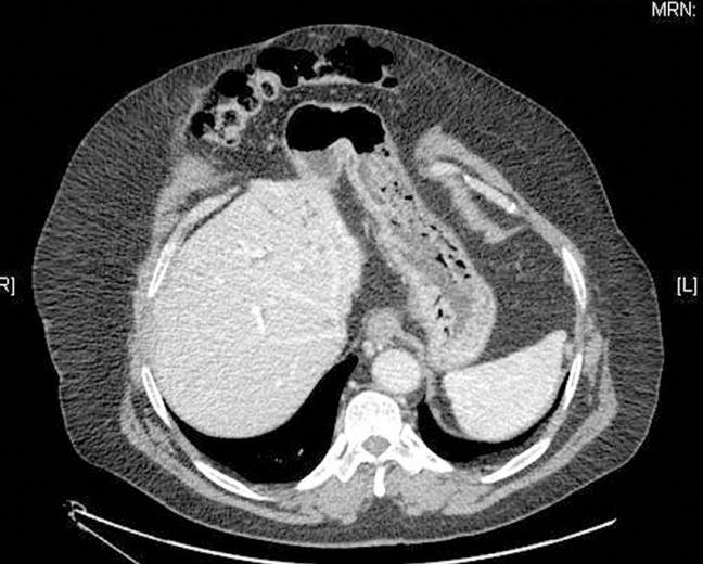
Figure 1 A CT scan showing severe obesity and umbilical laparocele.
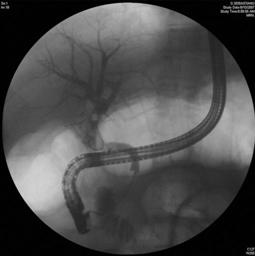
Figure 2 A first attempt of ERCP showing multiple stones and anastomotic stricture.
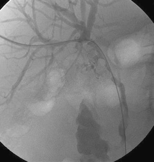
Figure 3 PTC with external - internal catheter placement.
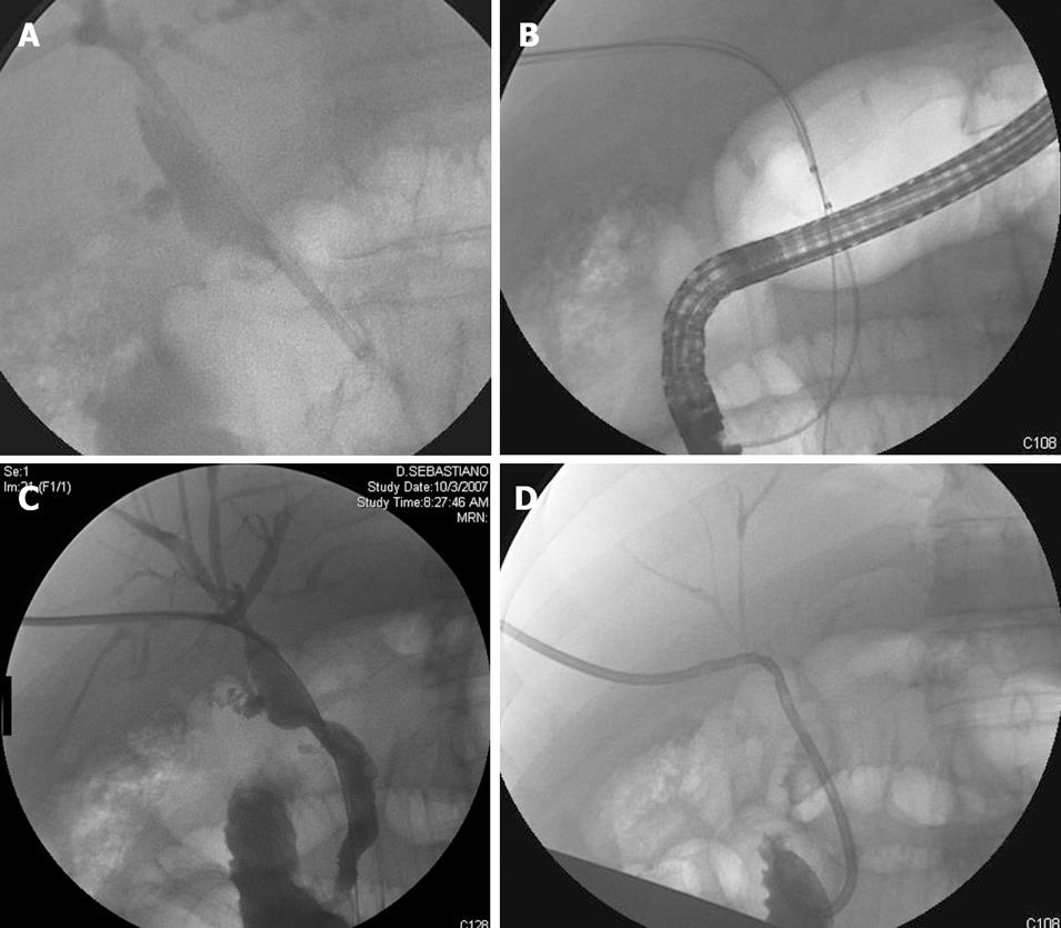
Figure 4 A: With a ballistic lithotripter, placed through the introductory, percutaneous lithotripsy was performed; B: Fragmented stones were removed with a Fogarty balloon; C: Normal Cholangiography after litotripsy and stenosis dilation; D: An external - internal catheter was placed.
- Citation: Pisa MD, Traina M, Miraglia R, Maruzzelli L, Volpes R, Piazza S, Luca A, Gridelli B. A case of biliary stones and anastomotic biliary stricture after liver transplant treated with the rendez - vous technique and electrokinetic lithotritor. World J Gastroenterol 2008; 14(18): 2920-2923
- URL: https://www.wjgnet.com/1007-9327/full/v14/i18/2920.htm
- DOI: https://dx.doi.org/10.3748/wjg.14.2920


