Copyright
©2008 The WJG Press and Baishideng.
World J Gastroenterol. Apr 14, 2008; 14(14): 2194-2199
Published online Apr 14, 2008. doi: 10.3748/wjg.14.2194
Published online Apr 14, 2008. doi: 10.3748/wjg.14.2194
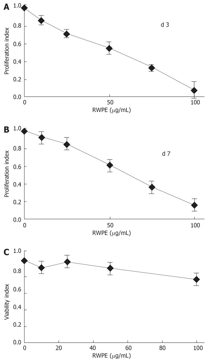
Figure 1 RWPE decreases myofibroblasts proliferation.
Myofibroblasts were grown for either three (A) or seven days (B) in the presence of the indicated concentrations of RWPE. Results are the mean ± SD of 4 independent experiments conducted in quadruplicate. The effect of RWPE was highly significant using ANOVA (P < 0.0001). In control experiments, myofibroblasts were exposed to RWPE for 24 h and the DNA content of the cell layer was measured (C). Results are expressed as the percentage of the values in treated cells as compared to cells treated with the solvent alone and are the mean ± SD of 3 independent experiments conducted in quadruplicate. There were no significant differences between conditions.
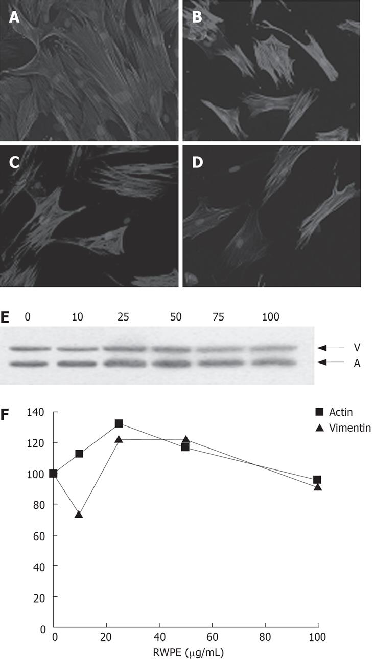
Figure 2 Effect of the RWPE on expression of alpha-smooth muscle actin.
A: Myofibroblasts were incubated for seven days in the absence of RWPE (A) or with 50 (B), 75 (C), or 100 (D) &mgr;g/mL RWPE. Aliquots of cell extracts grown in the same conditions were analyzed by Western blot (E) simultaneously for ASMA (A) and vimentin (V). F: The signals were quantified as described in Materials and Methods. The graph shows the mean of 2 separate experiments.
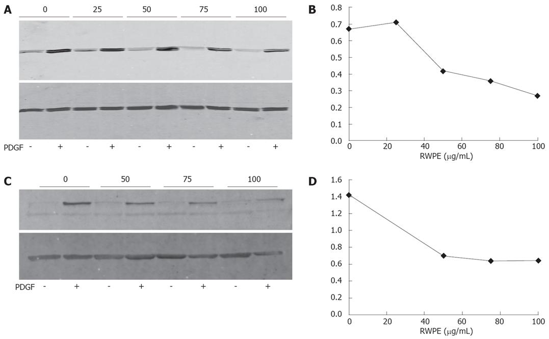
Figure 3 Effect of the RWPE on the phosphorylation of MAPK and Akt.
A: Myofibroblasts were pre-incubated for 1 h with the indicated concentrations of RWPE (in &mgr;g/mL) or solvent, then exposed for 10 min to 20 ng/mL PDGF-BB or buffer. Identical amounts of cell extracts were analyzed by Western blot with antibodies to phospho-ERK1/ERK2 (top panel) and to total ERKs (bottom panel). The picture is representative of 3 experiments; B: Quantitative analysis of the experiment shown in (A). The activation index refers to the ratio between the levels of phospho-ERK to those of total ERK; C: Same as in A except that the blot was labelled with an anti-phospho-Akt antibody (top panel) and an antibody to β-actin (bottom panel); D: Quantitative analysis of the experiment shown in (C). The activation index refers to the ratio between the levels of phospho-Akt to those of β-actin.
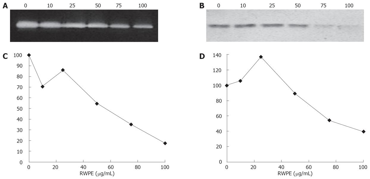
Figure 4 Effect of the RWPE on MMP-2 and TIMP-1 expression.
A: Myofibroblasts were incubated for 24 h with the indicated concentrations of RWPE (in &mgr;g/mL). Aliquots of conditioned medium normalized for the DNA content of the cell monolayers were analyzed on gelatin-containing gels (A). The white bands on the dark background indicate gelatinolytic activity. Other aliquots were analyzed by Western blot with an antibody against TIMP-1 (B). Another experiment gave similar results; C: Quantitative analysis of the experiments. MMP-2: Mean of duplicate samples; TIMP-1: Mean of 2 independent experiments.
- Citation: Neaud V, Rosenbaum J. A red wine polyphenolic extract reduces the activation phenotype of cultured human liver myofibroblasts. World J Gastroenterol 2008; 14(14): 2194-2199
- URL: https://www.wjgnet.com/1007-9327/full/v14/i14/2194.htm
- DOI: https://dx.doi.org/10.3748/wjg.14.2194


