Copyright
©2008 The WJG Press and Baishideng.
World J Gastroenterol. Apr 7, 2008; 14(13): 2115-2117
Published online Apr 7, 2008. doi: 10.3748/wjg.14.2115
Published online Apr 7, 2008. doi: 10.3748/wjg.14.2115
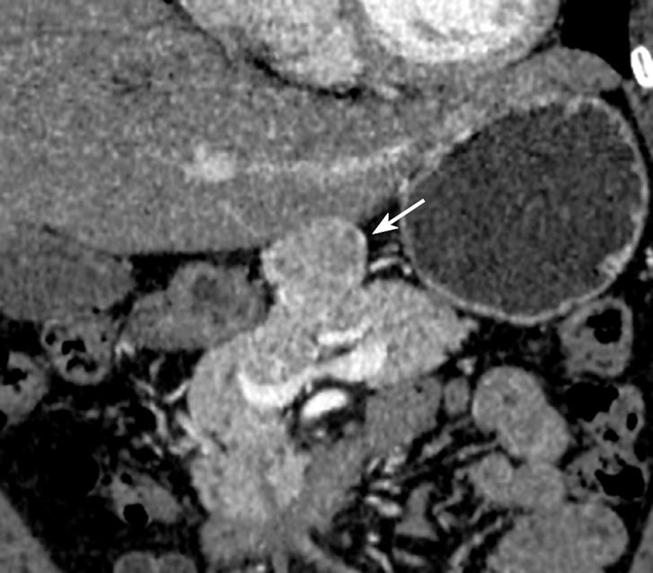
Figure 1 Pancreas dynamic CT scan showing a well-demarcated, arterial, enhancing mass measuring 3.
9 cm (arrow) just above the pancreatic body.
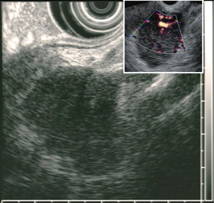
Figure 2 Endoscopic ultrasonography showing a well-delineated, homo-genous, elongated mass with a hypervascular appearance on Doppler (small box).
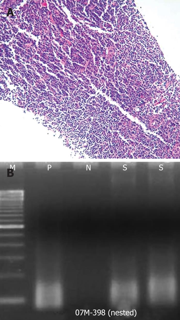
Figure 3 Microscopic examination of the EUS-TCB specimen showing expanded follicles replaced by a dense infiltration of small, mature, lymphocytes devoid of follicular center cells (A) (× 200) and PCR study for IgH gene rearrangement showing a polyclonal pattern (B).
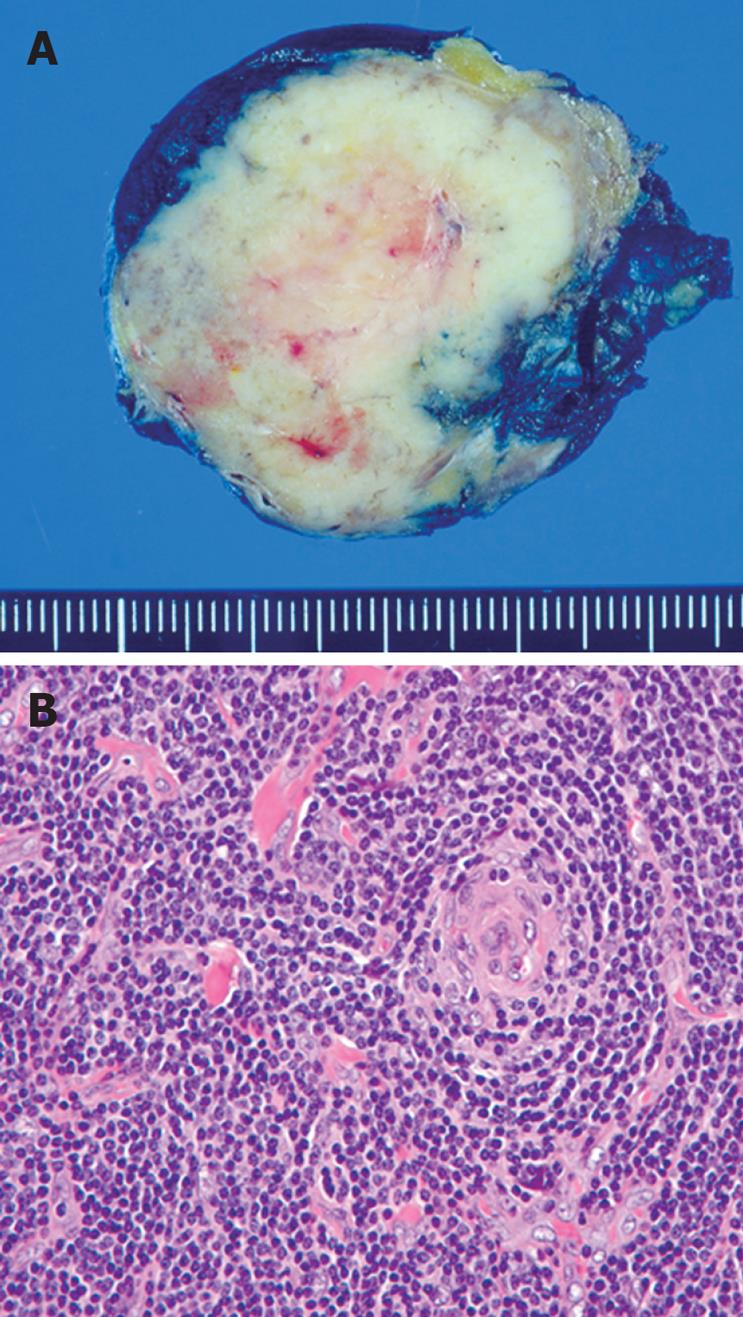
Figure 4 Surgical specimen showing creamy white cut surface of the resected tumor with trabeculation and numerous, small blood vessels (A) and lymph node demonstrating burnt-out, atrophic follicles with depletion of follicle center cells and residual, follicular, dendritic cells and surrounding, concentric layers of small lymphocytes (B).
Radial penetrating blood vessels are also seen (× 400).
- Citation: Rhee KH, Lee SS, Huh JR. Endoscopic ultrasonography-guided trucut biopsy for the preoperative diagnosis of peripancreatic castleman’s disease: A case report. World J Gastroenterol 2008; 14(13): 2115-2117
- URL: https://www.wjgnet.com/1007-9327/full/v14/i13/2115.htm
- DOI: https://dx.doi.org/10.3748/wjg.14.2115


