Copyright
©2008 The WJG Press and Baishideng.
World J Gastroenterol. Mar 28, 2008; 14(12): 1958-1960
Published online Mar 28, 2008. doi: 10.3748/wjg.14.1958
Published online Mar 28, 2008. doi: 10.3748/wjg.14.1958
Figure 1 Chronological changes in US findings.
A: Initial US showed a low-echoic mass, 7 mm in diameter, in the pancreatic body (arrow); B: After 10 mo, the diameter of the low-echoic mass had increased to 9 mm (arrow); C: After 22 mo, the diameter of the low-echoic mass had increased to 13 mm (arrow); D: After 22 mo, a grape-like cyst distal to the low-echoic mass was detected for the first time (arrow).
Figure 2 Chronological changes in EUS findings.
A: Initial EUS demonstrated an extremely low-echoic mass with partial posterior echo enhancement, 7 mm in diameter, in the pancreatic body (arrow); B: After 14 mo, the diameter of the low-echoic mass had increased to 9 mm (arrow); C: After 22 mo, the diameter of the low-echoic mass had increased to 13 mm (arrow).
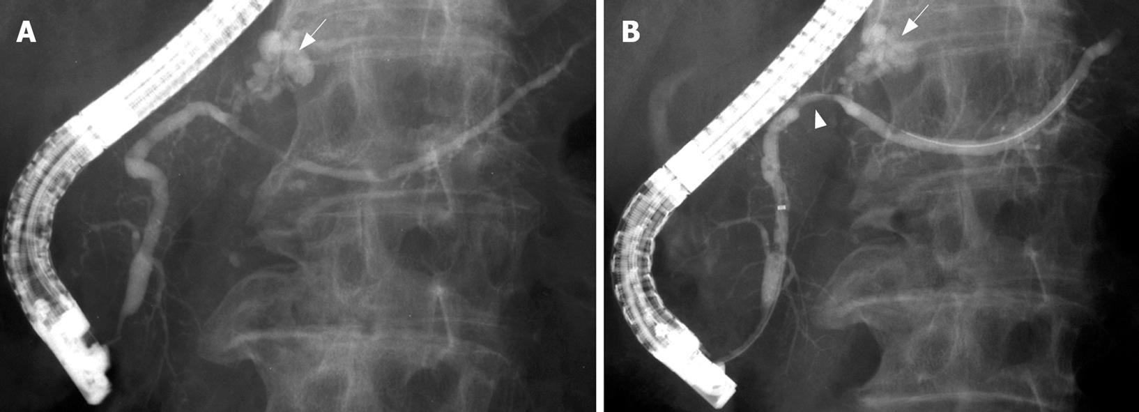
Figure 3 Chronological changes in ERP findings.
A: Initial ERP revealed a dilated branch duct in the pancreatic body (arrow), and the MPD was mildly dilated by mucus; B: After 22 mo, the dilated branch duct did not show any marked change (arrow). Proximal to it, the MPD was slightly compressed (arrow head).
Figure 4 Histopathological findings.
A: The mass is composed of moderately differentiated tubular adenocarcinoma with desmoplastic fibrosis, resulting in a diagnosis of IDC (HE, original magnification × 20); B: The tumor mass includes macroscopic cystic components (*) lined by normal ductal epithelium, suggestive of retention cysts in carcinoma (HE, original magnification × 1); C: The lining of the dilated branch duct is composed of low-papillary columnar cells with copious intracellular mucus, resulting in a diagnosis of branch duct type intraductal papillary-mucinous adenoma with mild atypia (HE, × 40).
- Citation: Hisa T, Ohkubo H, Shiozawa S, Ishigame H, Takamatsu M, Furutake M, Nobukawa B, Suda K. Growth process of small pancreatic carcinoma: A case report with imaging observation for 22 months. World J Gastroenterol 2008; 14(12): 1958-1960
- URL: https://www.wjgnet.com/1007-9327/full/v14/i12/1958.htm
- DOI: https://dx.doi.org/10.3748/wjg.14.1958


