Copyright
©2008 The WJG Press and Baishideng.
World J Gastroenterol. Mar 28, 2008; 14(12): 1946-1948
Published online Mar 28, 2008. doi: 10.3748/wjg.14.1946
Published online Mar 28, 2008. doi: 10.3748/wjg.14.1946
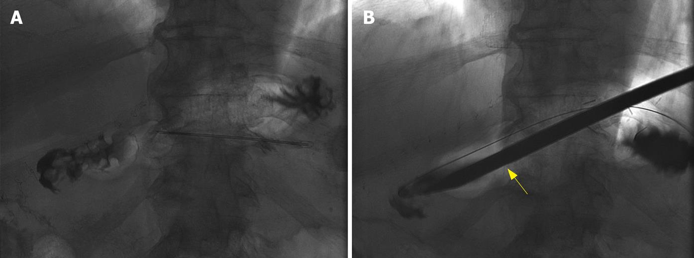
Figure 1 Fluoroscopic images of percutaneous endoscopy.
A: Inflated bypassed stomach with air; B: Trochar (arrow) used to dilate the percutaneous track.
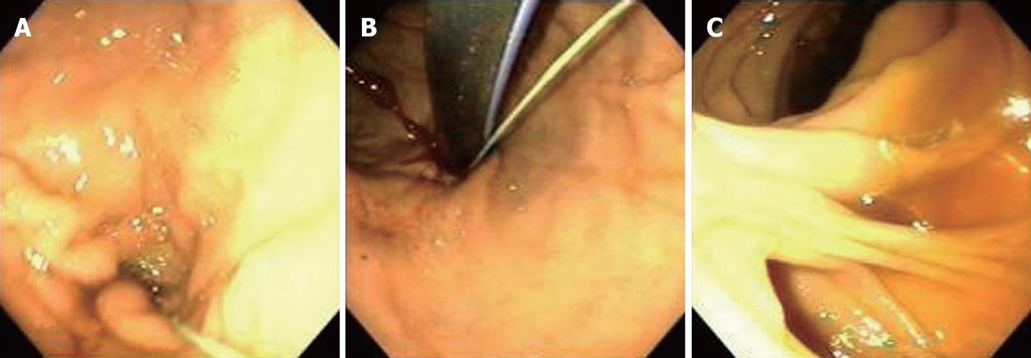
Figure 2 Endoscopic images by percutaneous endoscopy.
A: Antegrade views of bypassed stomach; B: Retrograde views of bypassed stomach; C: Roux-en-Y anastomosis.
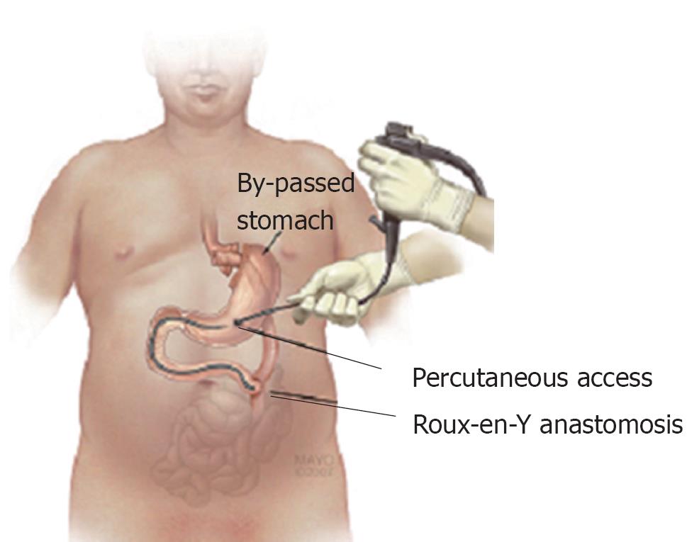
Figure 3 Illustration of the percutaneous endoscopy.
- Citation: Gill KR, McKinney JM, Stark ME, Bouras EP. Investigation of the excluded stomach after Roux-en-Y gastric bypass: The role of percutaneous endoscopy. World J Gastroenterol 2008; 14(12): 1946-1948
- URL: https://www.wjgnet.com/1007-9327/full/v14/i12/1946.htm
- DOI: https://dx.doi.org/10.3748/wjg.14.1946


