Copyright
©2008 The WJG Press and Baishideng.
World J Gastroenterol. Mar 28, 2008; 14(12): 1842-1850
Published online Mar 28, 2008. doi: 10.3748/wjg.14.1842
Published online Mar 28, 2008. doi: 10.3748/wjg.14.1842
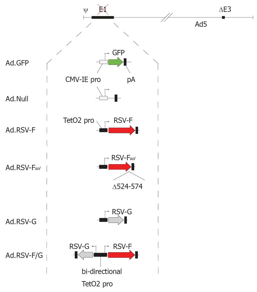
Figure 1 Adenovirus vector design.
All adenovirus vectors used in this study are E1- and E3-deleted and Ad5-based. The vector Ad.GFP encodes green fluorescent protein and the vector Ad.Null served as an adenovirus vector control. The RSV glycoprotein encoding vectors carried the transgene under the transcriptional control of the doxycycline-repressible TetO2 promoter. The vector RSV-Fsol encodes a soluble form of the RSV fusion protein F without the transmembrane domain and cytoplasmic tail (Δ524-574).
Figure 2 Intratumoral expression of RSV-F or RSV-F/G induces an anti-neoplastic effect.
In a bilateral subcutaneous syngeneic MC38 or Colon26 colon cancer model, the indicated adenovirus vectors were inoculated on d 0 and d 2 into the tumor on the right flank. No viral vectors were inoculated into the tumor on the left flank. A: The volume of the tumor on the right flank was measured at d 28 and presented as box-and-whisker plots, showing minimum, 25th percentile, median, 75th percentile, and maximum tumor volume; B: The volume of the tumor on the left flank, which did not receive direct viral vector injections, was measured at d 28 and the volume reduction relative to Ad.Null-treated control animals is presented as bar graphs (mean ± SD).
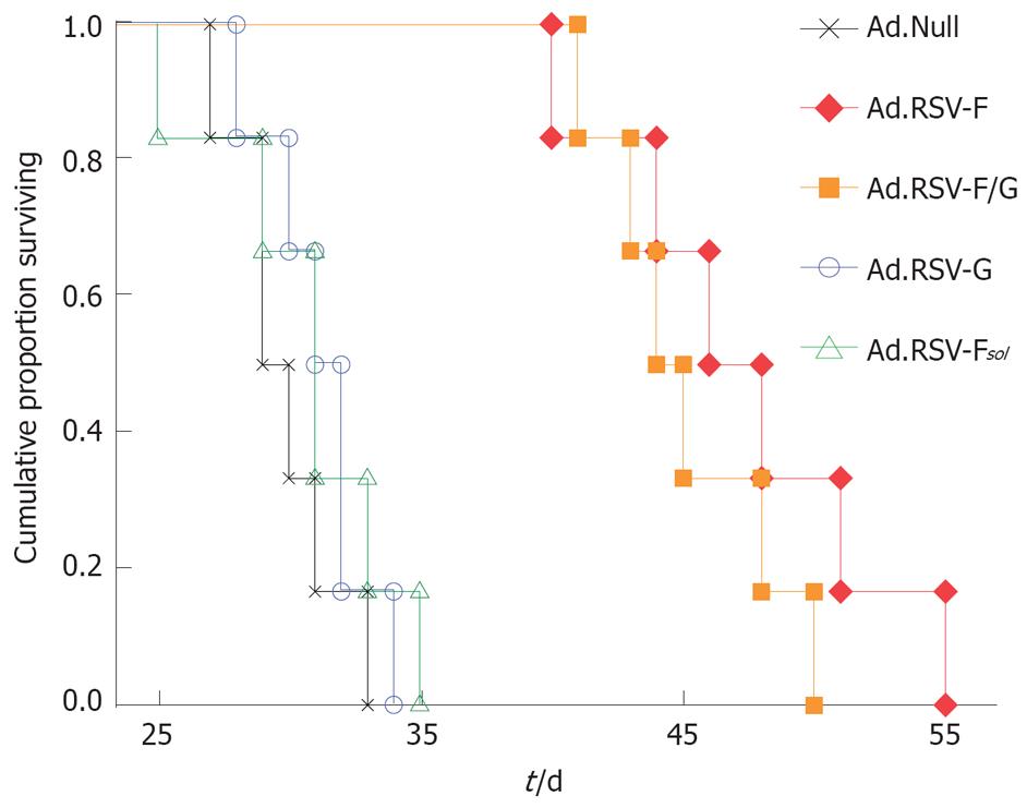
Figure 3 Kaplan-Meier survival analysis.
In the bilateral subcutaneous syngeneic MC38 colon cancer model described in Figure 2, one tumor was treated with the indicated adenoviral vectors, and survival time was monitored up to d 55.
Figure 4 Fluorescence micrographs.
MC38 cells were transduced in vitro with indicated RSV glycoprotein encoding adenoviruses and an adenovirus encoding GFP. Transduction with Ad.GFP enhanced the visibility of syncytia. Representative pictures 48 h after transduction are shown (× 200). Similar data were obtained with the Colon26 cells (data not shown).
Figure 5 Analysis of apoptosis.
A: MC38 or Colon26 cells were transduced in vitro with indicated adenovirus vectors and early apoptotic events were analyzed by flow cytometric measurements of phosphatidylserine translocation to the outer membrane by annexin V binding; B: Because apoptosis is essentially executed by proteases of the caspase family, we analyzed caspase-3/7 activity. Relative caspase activities are given as means ± SD of three independent experiments; C: To measure mitochondrial alterations we determined the mitochondrial membrane potential ΔΨm. Ad.Null served as a control.
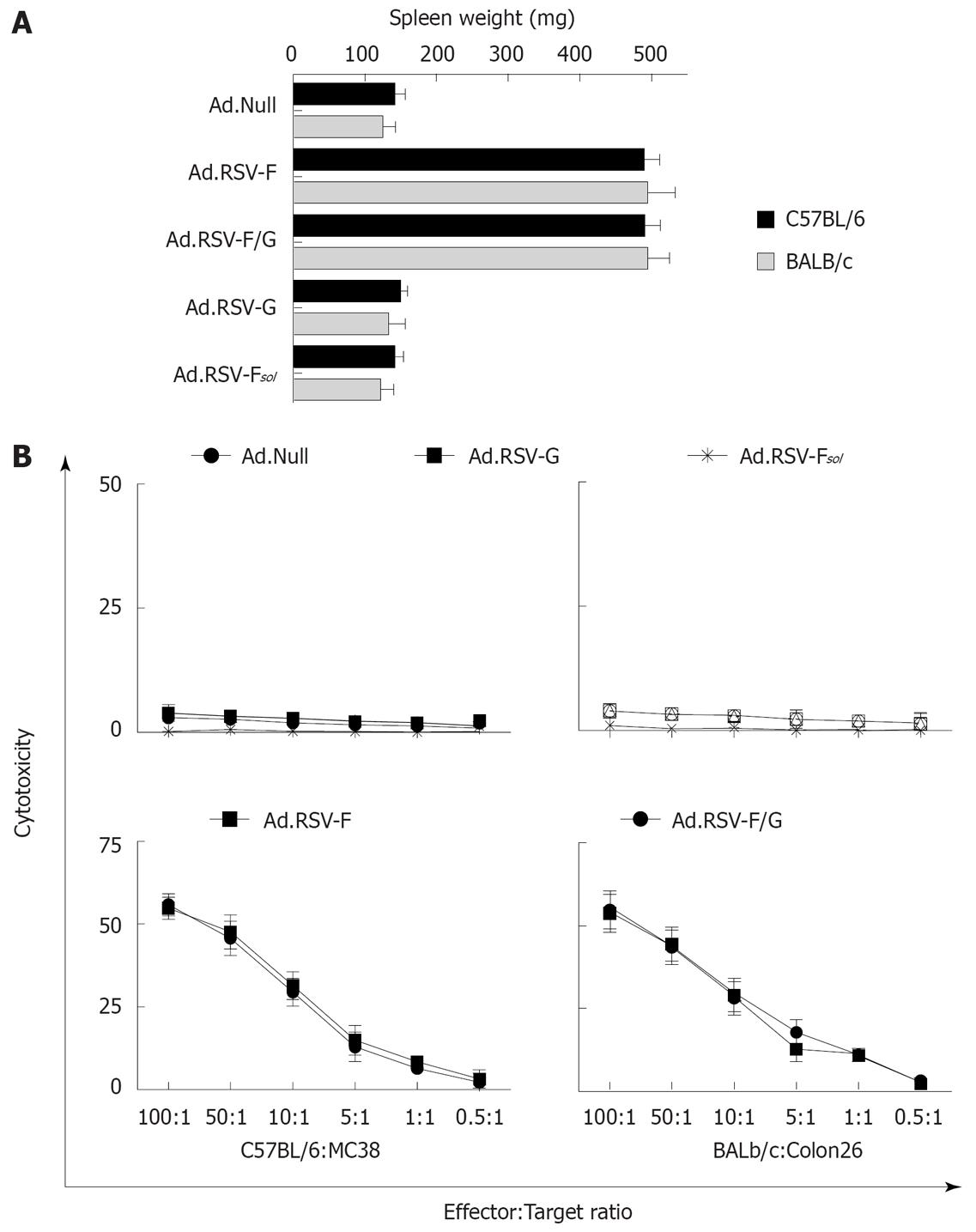
Figure 6 Effects of indicated treatments on the spleen weight and cytotoxic T cell induction.
A: In the bilateral subcutaneous syngeneic MC38 and Colon26 tumor model described in Figure 2, animals were euthanized at day 28 and spleen weight was determined (mean ± SD); B: In addition, we determined the cytotoxic activity of spleen-derived T cells against target MC38 or Colon26 tumor cells in these animals using an LDH release assay. Data of all animals were expressed as the average percentage of specific LDH release from three independent experiments (mean ± SD).
- Citation: Hoffmann D, Grunwald T, Bayer W, Wildner O. Immune-mediated anti-neoplastic effect of intratumoral RSV envelope glycoprotein expression is related to apoptotic death of tumor cells but not to the size of syncytia. World J Gastroenterol 2008; 14(12): 1842-1850
- URL: https://www.wjgnet.com/1007-9327/full/v14/i12/1842.htm
- DOI: https://dx.doi.org/10.3748/wjg.14.1842


