Copyright
©2008 The WJG Press and Baishideng.
World J Gastroenterol. Mar 28, 2008; 14(12): 1836-1841
Published online Mar 28, 2008. doi: 10.3748/wjg.14.1836
Published online Mar 28, 2008. doi: 10.3748/wjg.14.1836
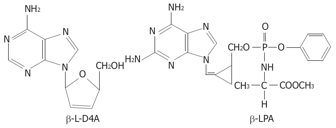
Figure 1 Structures of β-L-D4A and β-LPA.
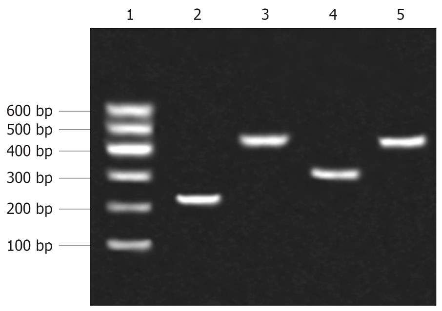
Figure 2 The HBV promoters obtained from PCR were separated by agarose gel electrophoresis.
Lane 1: Marker; Lane 2: preSp; Lane 3: Sp; Lane 4: Cp; Lane 5: Xp.
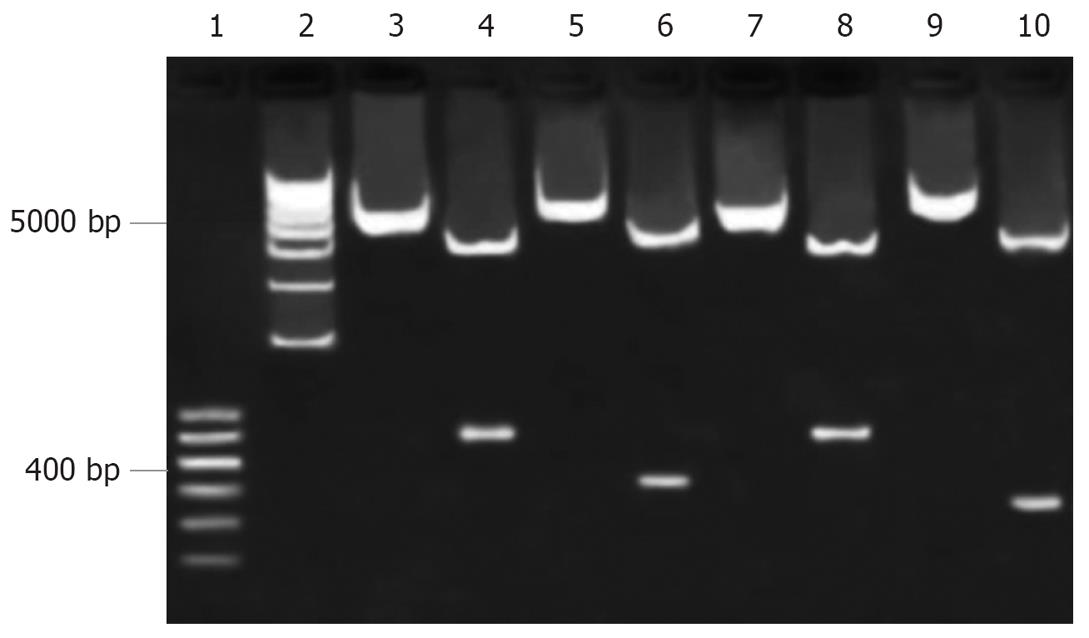
Figure 3 Restriction analysis of recombinant vectors.
Lanes 1, 2: Marker; Lanes 3, 4: pEGFP-Sp before and after being digested by Acc65Iand AgeI; Lanes 5, 6: pEGFP-Cp before and after being digested by Acc65Iand AgeI; Lanes 7, 8: pEGFP-Xp before and after being digested by Acc65Iand AgeI; Lanes 9, 10: pEGFP-preSp before and after being digested by Acc65Iand AgeI.
Figure 4 Forty-eight hours after transfection, EGFP positive cells were detected by fluorescence microscopy (× 100).
A: HepG2 cells transfected with pEGFP-Sp; B: HepG2 cells transfected with pEGFP-Cp; C: HepG2 cells transfected with pEGFP-Xp; D: HepG2 cells transfected with pEGFP-preSp.
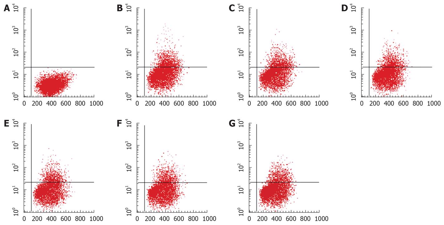
Figure 5 β-L-D4A inhibited the expression of EGFP in HepG2 cells transfected with pEGFP-Sp, as determined by FACS analysis.
A: HepG2 cells not transfected with pEGFP-Sp, not treated with drug, the percentage of EGFP-positive cells was 0; B: HepG2 cells transfected with pEGFP-Sp, not treated with drug, the percentage was 21.42%; C: HepG2 cells transfected with pEGFP-Sp, treated with Lamivudine at 1 &mgr;mol/L, the percentage was 21.14%; D-G: HepG2 cells transfected with pEGFP-Sp, treated with β-L-D4A at various concentrations (0.08, 0.4, 2, 10 &mgr;mol/L), the percentages were 18.76%, 17.31%, 15.53%, 13.65% respectively.
- Citation: He XX, Lin JS, Chang Y, Zhang YH, Li Y, Wang XY, Xu D, Cheng XM. Effects of two novel nucleoside analogues on different hepatitis B virus promoters. World J Gastroenterol 2008; 14(12): 1836-1841
- URL: https://www.wjgnet.com/1007-9327/full/v14/i12/1836.htm
- DOI: https://dx.doi.org/10.3748/wjg.14.1836


