Copyright
©2008 The WJG Press and Baishideng.
World J Gastroenterol. Mar 21, 2008; 14(11): 1710-1719
Published online Mar 21, 2008. doi: 10.3748/wjg.14.1710
Published online Mar 21, 2008. doi: 10.3748/wjg.14.1710
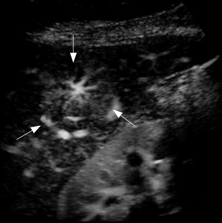
Figure 1 Contrast-enhanced harmonic imaging with Sonazoid in focal nodular hyperplasia (FNH).
The centrifugal blood flow appearance like “spoke-wheel sign” was clearly demonstrated in the center of the nodules (arrows).
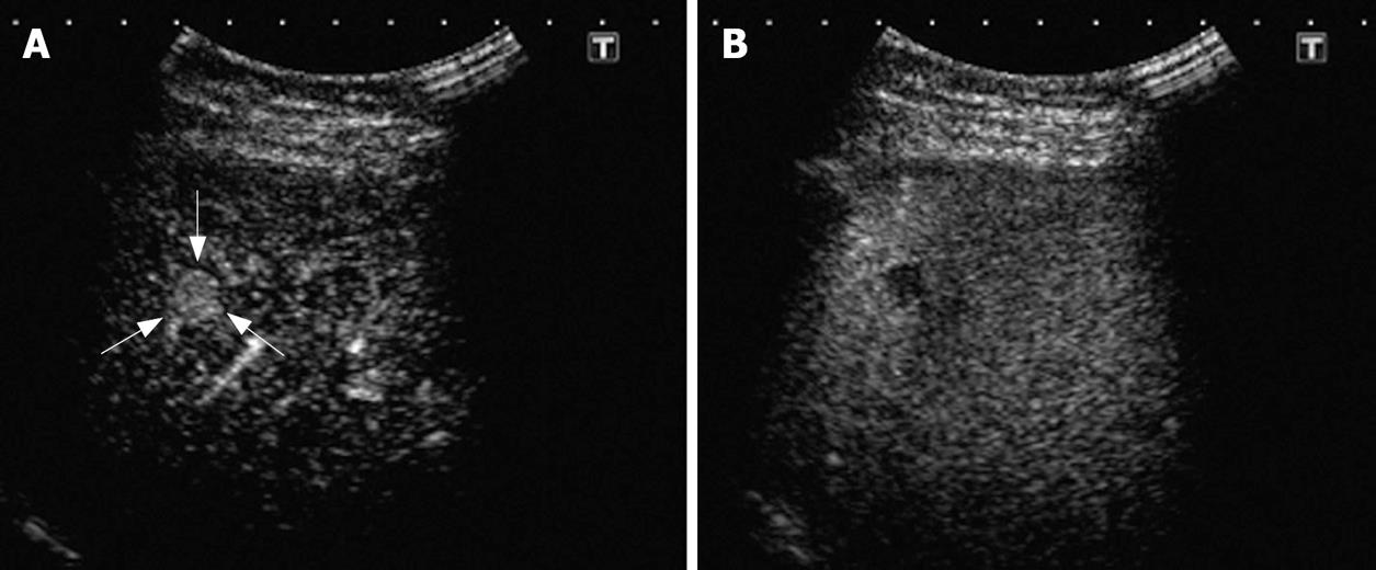
Figure 2 Contrast-enhanced harmonic imaging with Sonazoid in small HCC (9.
8 mm, arrows). A: Early-phase image (22 s after the injection); B: Late-phase image (10 min after the injection). The early-phase image showed positive enhancement and the late-phase image showed negative enhancement in the nodule. These findings could easily diagnose this lesion as HCC.
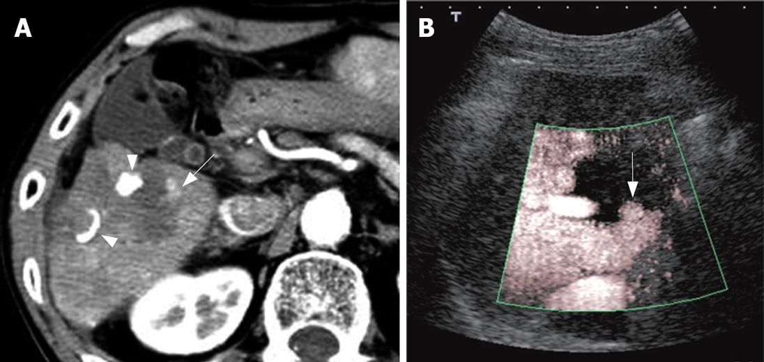
Figure 3 Assessment of therapeutic response after PEI for HCC.
A: Contrast-enhanced CT with dynamic study; B: Contrast-enhanced US (Advanced Dynamic Flow with Levovist). Contrast-enhanced CT showed enhancement appearance which needed additional treatment within the treated area (arrow), and contrast-enhanced US could demonstrate a similar finding. Arrow heads: Lipiodol.
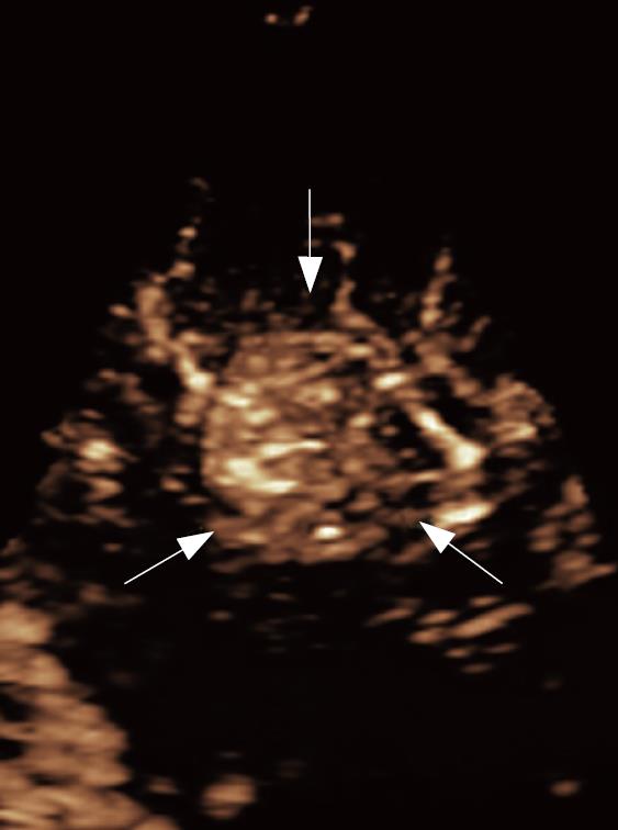
Figure 4 Real-time three-dimensional imaging of HCC (contrast-enhanced LIVE 3D with Sonazoid, iu22, Philips).
Abundant tumor vessels were dramatically demonstrated in the HCC nodule. (Arrows: HCC nodule).
- Citation: Maruyama H, Yoshikawa M, Yokosuka O. Current role of ultrasound for the management of hepatocellular carcinoma. World J Gastroenterol 2008; 14(11): 1710-1719
- URL: https://www.wjgnet.com/1007-9327/full/v14/i11/1710.htm
- DOI: https://dx.doi.org/10.3748/wjg.14.1710


