Copyright
©2008 The WJG Press and Baishideng.
World J Gastroenterol. Mar 14, 2008; 14(10): 1633-1635
Published online Mar 14, 2008. doi: 10.3748/wjg.14.1633
Published online Mar 14, 2008. doi: 10.3748/wjg.14.1633
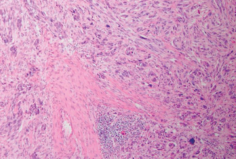
Figure 1 Histopathological examination of a biopsy taken at rectosigmoidoscopy showed the tumour to be a leiomyosarcoma (HE, × 100).
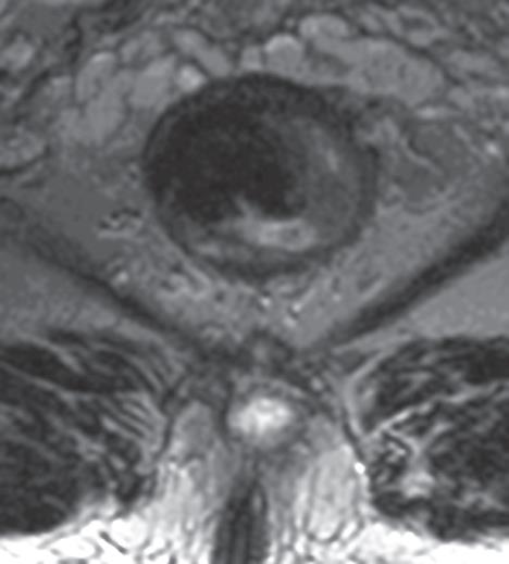
Figure 2 MRI-rectum showing a tumour just behind the anal verge.
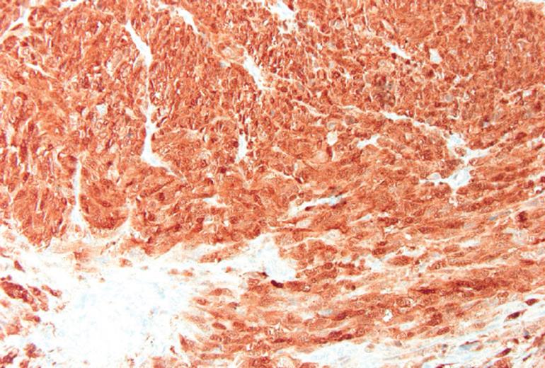
Figure 3 Histopathological examination of the abdomino-perineal resection (S-100 Positive, x 200).
S-100 staining performed with an automatic system (Benchmark XT, Ventana Systems). Diffuse expression of S-100 staining typical for melanoma. In leiomyosarcoma, S-100 is never expressed.
- Citation: Schaik PV, Ernst M, Meijer H, Bosscha K. Melanoma of the rectum: A rare entity. World J Gastroenterol 2008; 14(10): 1633-1635
- URL: https://www.wjgnet.com/1007-9327/full/v14/i10/1633.htm
- DOI: https://dx.doi.org/10.3748/wjg.14.1633


