Copyright
©2008 The WJG Press and Baishideng.
World J Gastroenterol. Jan 7, 2008; 14(1): 143-145
Published online Jan 7, 2008. doi: 10.3748/wjg.14.143
Published online Jan 7, 2008. doi: 10.3748/wjg.14.143
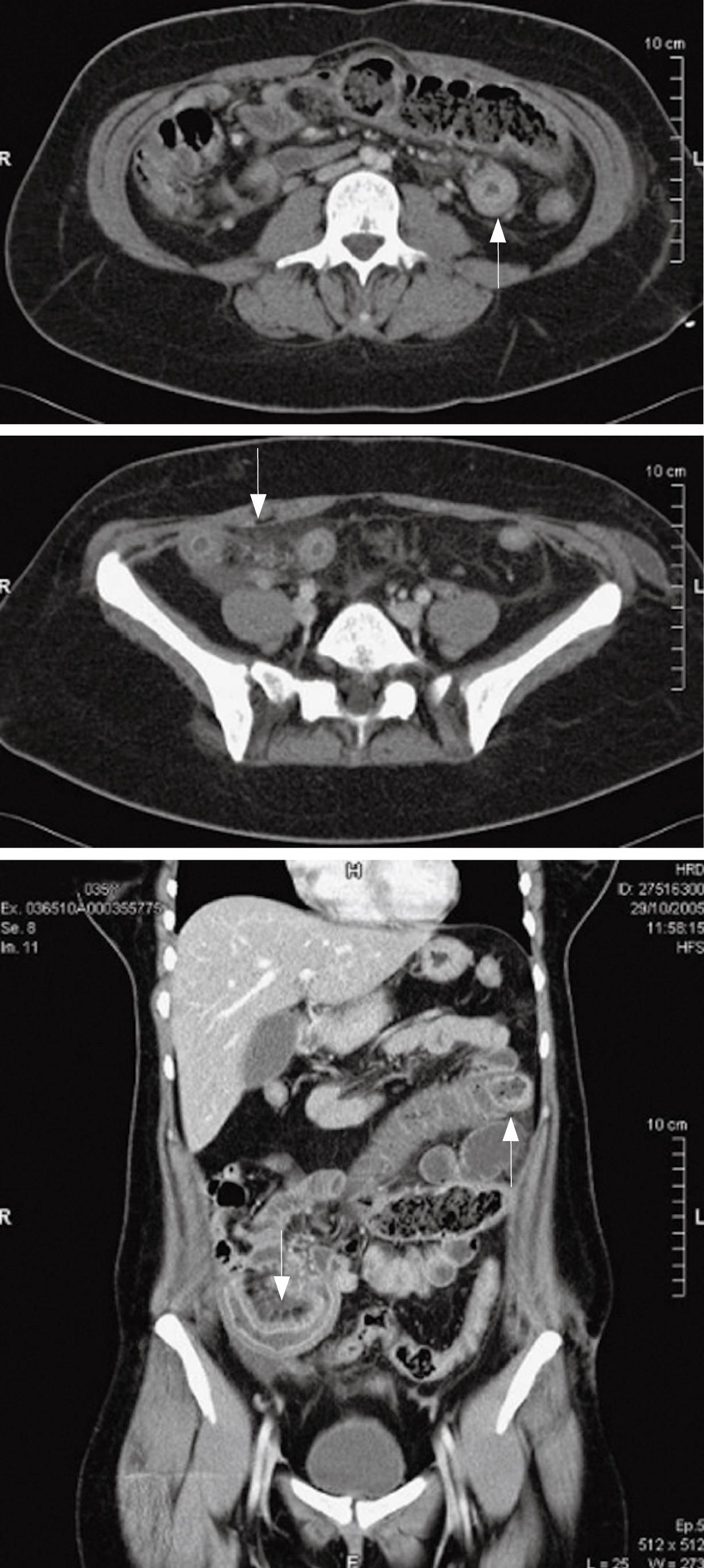
Figure 1 A: Transversal abdominal computed tomography, demonstrating two thickened bowel loops with mesenteric haziness (arrows); B: Frontal abdominal computed tomography, demonstrating two thickened bowel loops with mesenteric haziness (arrows).
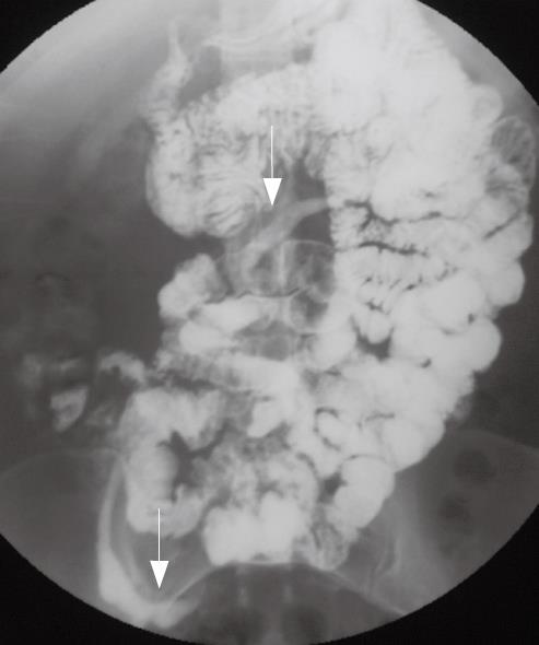
Figure 2 Abdominal enteroclysis showing two intestinal strictures, one proximal, the other distal (arrows).
Figure 3 Preoperative picture showing two thickened, fibrotic small bowel segments (arrows) in front of a scarred mesenteric tear.
Figure 4 Inflammation with mucosal ulceration.
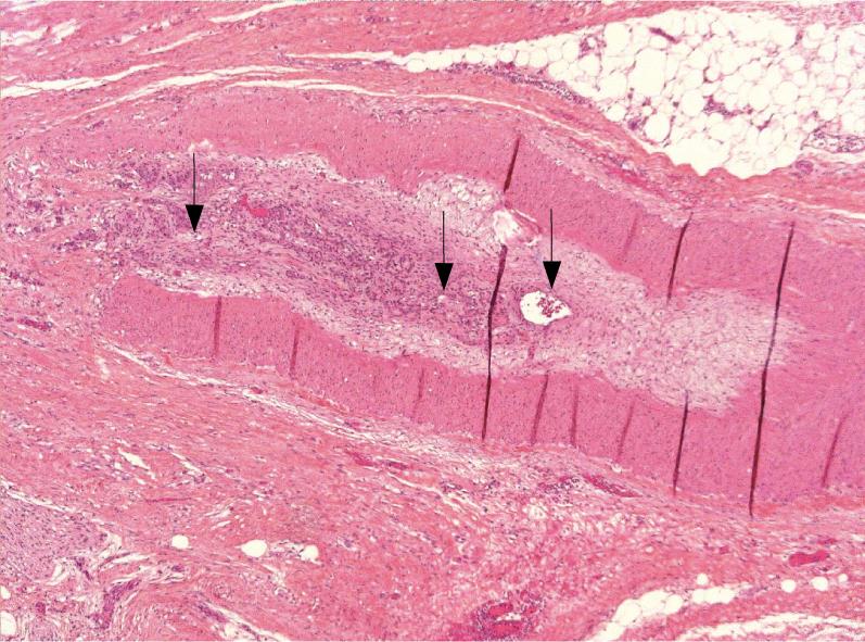
Figure 5 Microscopic section of healed mesenteric injury showing vascular chronic lesion with a repermeabilised venous thrombosis (arrows).
- Citation: Bougard V, Avisse C, Patey M, Germain D, Levy-Chazal N, Delattre JF. Double ischemic ileal stenosis secondary to mesenteric injury after blunt abdominal trauma. World J Gastroenterol 2008; 14(1): 143-145
- URL: https://www.wjgnet.com/1007-9327/full/v14/i1/143.htm
- DOI: https://dx.doi.org/10.3748/wjg.14.143


