Copyright
©2008 The WJG Press and Baishideng.
World J Gastroenterol. Jan 7, 2008; 14(1): 101-107
Published online Jan 7, 2008. doi: 10.3748/wjg.14.101
Published online Jan 7, 2008. doi: 10.3748/wjg.14.101
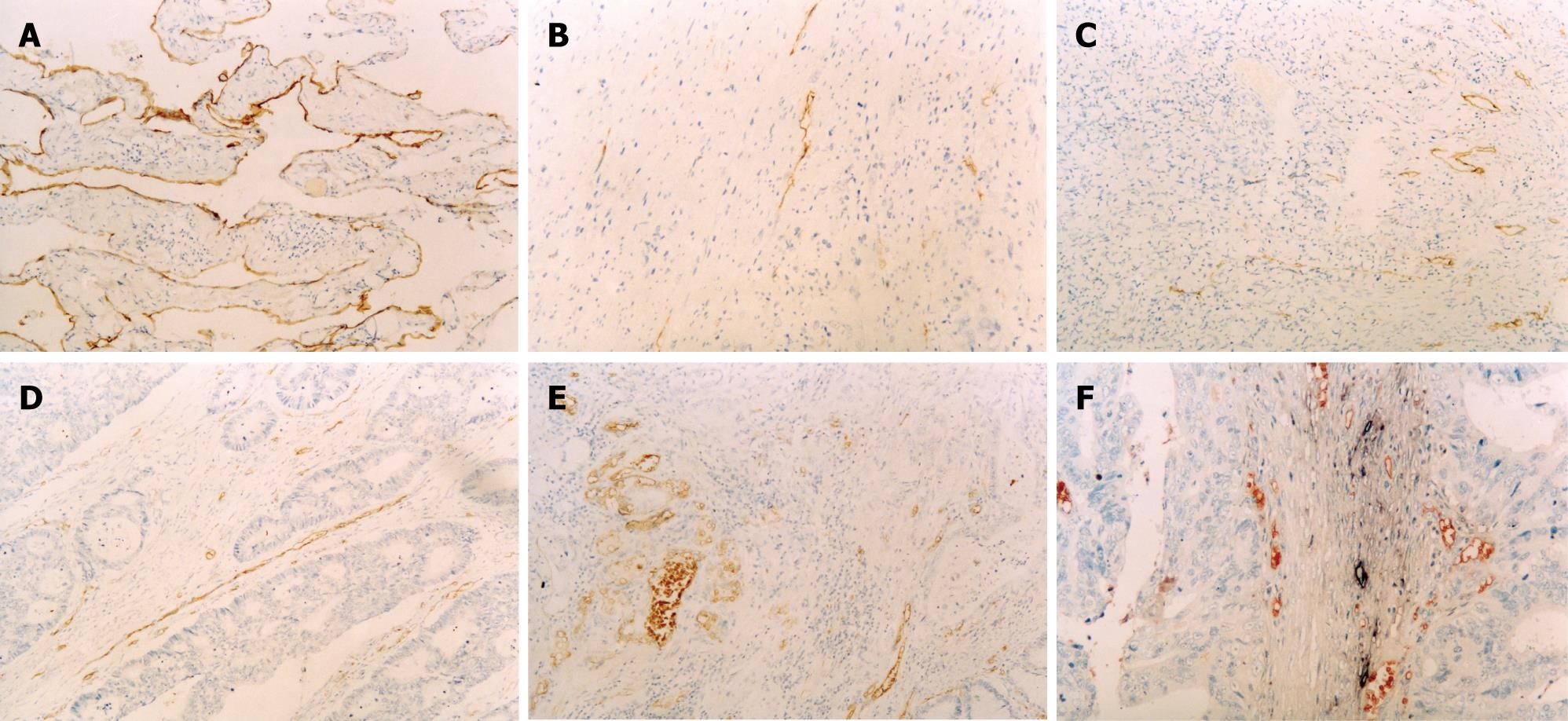
Figure 1 Immunohistochemical stainings of D2-40 (A, B, C × 100), vWF (D, E × 100) and double labeling immunohistochemistry (F × 200, red: Blood vessels labled by vWF; amethyst: Lymphatic vessels by D2-40).
A: Lymphangioma (positive control); B-F: Colorectal carcinoma.
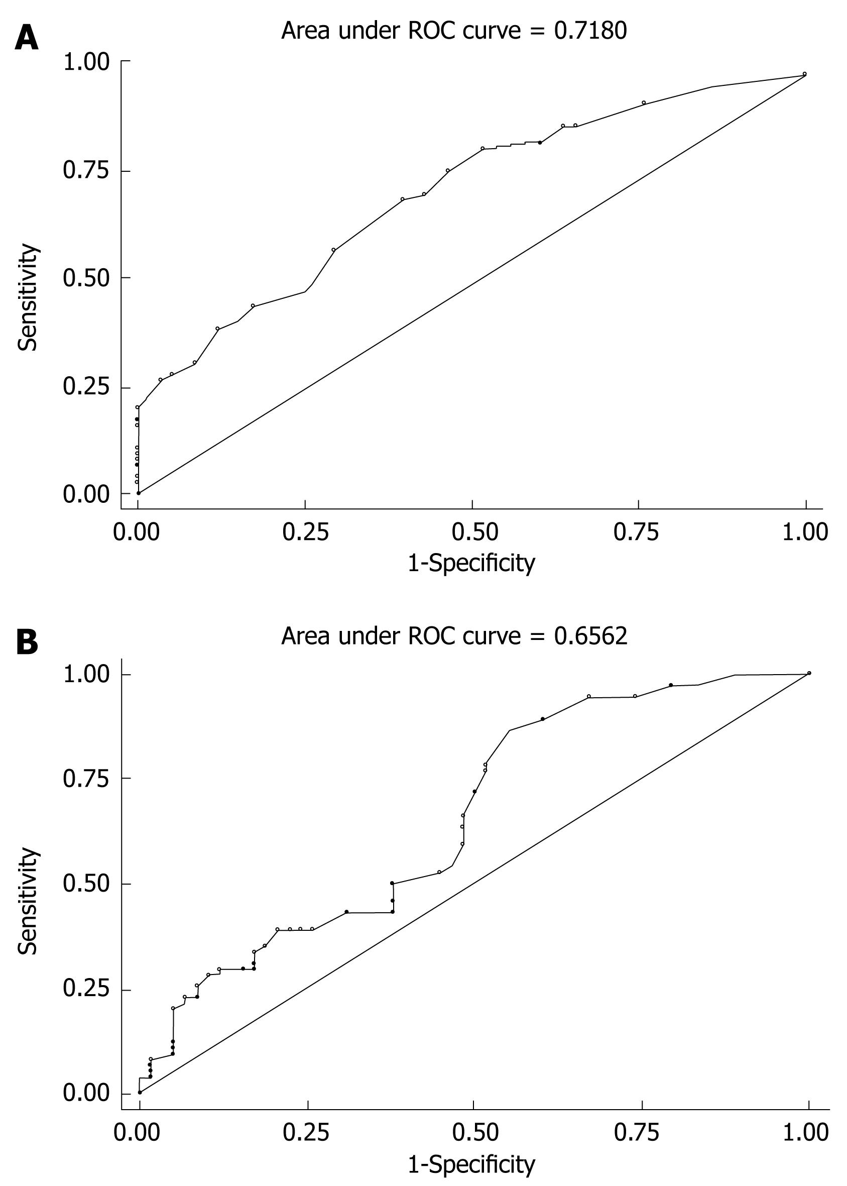
Figure 2 ROC curve of LVD (A) and MVD (B).
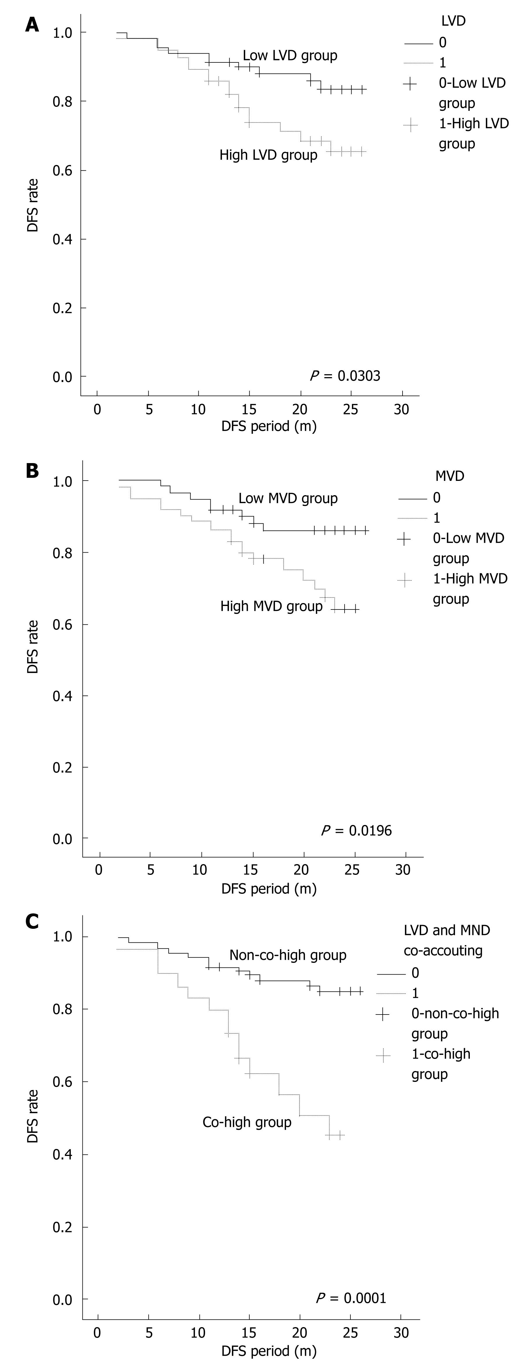
Figure 3 Survival curve of LVD (A), MVD (B) and co-accounting of LVD and MVD (C).
- Citation: Yan G, Zhou XY, Cai SJ, Zhang GH, Peng JJ, Du X. Lymphangiogenic and angiogenic microvessel density in human primary sporadic colorectal carcinoma. World J Gastroenterol 2008; 14(1): 101-107
- URL: https://www.wjgnet.com/1007-9327/full/v14/i1/101.htm
- DOI: https://dx.doi.org/10.3748/wjg.14.101


