Copyright
©2007 Baishideng Publishing Group Co.
World J Gastroenterol. Mar 7, 2007; 13(9): 1466-1470
Published online Mar 7, 2007. doi: 10.3748/wjg.v13.i9.1466
Published online Mar 7, 2007. doi: 10.3748/wjg.v13.i9.1466
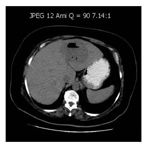
Figure 1 Contrast en-hanced CT scan showing a low-density area with gas and fluid, measuring approximately 8.
5 cm x 7.0 cm, consistent with left-sided intra-hepatic abscess.
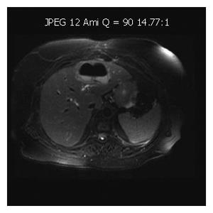
Figure 2 Abdominal RM demonstrating a large collection with gas and fluid.
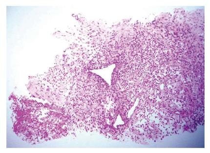
Figure 3 Biopsy of the liver abscess showing fibrosis, fibrin and acute inflammatory cells, consistent with abscess wall (HE).
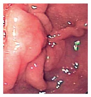
Figure 4 Upper GI endoscopy revealing a thickened gastric fold (pre-pyloric).
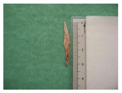
Figure 5 Removed foreign body (chicken bone, with 3.
3 cm x 0.5 cm).
- Citation: Santos SA, Alberto SC, Cruz E, Pires E, Figueira T, Coimbra &, Estevez J, Oliveira M, Novais L, Deus JR. Hepatic abscess induced by foreign body: Case report and literature review. World J Gastroenterol 2007; 13(9): 1466-1470
- URL: https://www.wjgnet.com/1007-9327/full/v13/i9/1466.htm
- DOI: https://dx.doi.org/10.3748/wjg.v13.i9.1466


