Copyright
©2007 Baishideng Publishing Group Co.
World J Gastroenterol. Feb 28, 2007; 13(8): 1299-1302
Published online Feb 28, 2007. doi: 10.3748/wjg.v13.i8.1299
Published online Feb 28, 2007. doi: 10.3748/wjg.v13.i8.1299
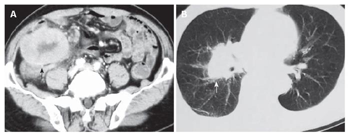
Figure 1 Computed tomography showing a large soft-tissue mass (black arrow) in the right lumbar region and iliac fossa (A) and a mass (white arrow) on the hilum of the right lung (B).
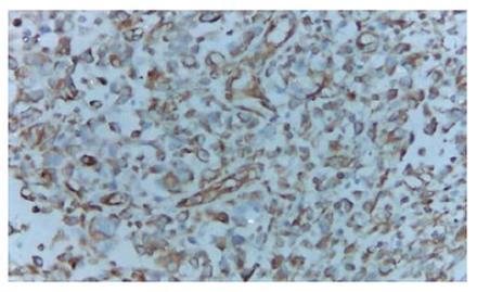
Figure 2 Tumor cells showing positive staining for vimentin (EnVision × 200).
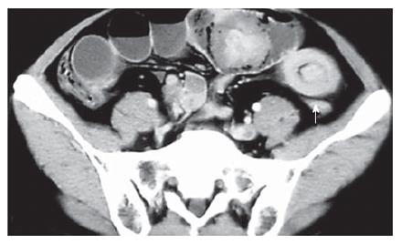
Figure 3 Computed tomography showing features of multiple intussusceptions in the small intestine (white arrow).
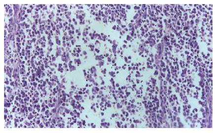
Figure 4 Histology revealing tumor cells consisting of fibroblast-like cells, histiocyte-like cells and pleomorphic giant cells (HE, × 40).
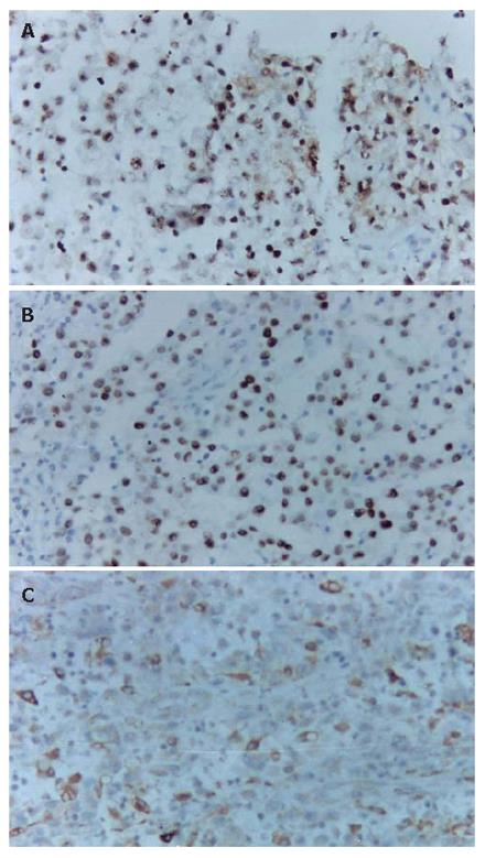
Figure 5 Tumor cells showing positive staining for Ki67 (A), P53 (B), and KP-1 (C) (EnVision × 200).
- Citation: Fu DL, Yang F, Maskay A, Long J, Jin C, Yu XJ, Xu J, Zhou ZW, Ni QX. Primary intestinal malignant fibrous histiocytoma: Two case reports. World J Gastroenterol 2007; 13(8): 1299-1302
- URL: https://www.wjgnet.com/1007-9327/full/v13/i8/1299.htm
- DOI: https://dx.doi.org/10.3748/wjg.v13.i8.1299


