Copyright
©2007 Baishideng Publishing Group Co.
World J Gastroenterol. Feb 28, 2007; 13(8): 1279-1281
Published online Feb 28, 2007. doi: 10.3748/wjg.v13.i8.1279
Published online Feb 28, 2007. doi: 10.3748/wjg.v13.i8.1279
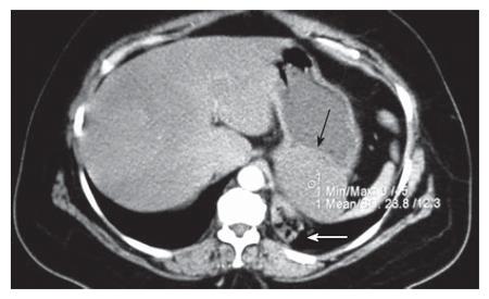
Figure 1 Abdominal CT scan demonstrating a cystic lesion attached to the posterior gastric wall (black arrow) and a pulmonary sequestration in the left pulmonary base (white arrow).
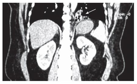
Figure 2 Coronary CT plate showing the pulmonary sequestration of the basal segment of the left lower lobe (arrow).
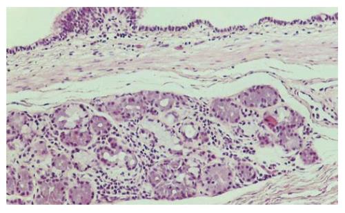
Figure 3 Histological section of the cyst wall lined by a respiratory-type epithelium and containing seromucinous glands (HE, x 100).
- Citation: Theodosopoulos T, Marinis A, Karapanos K, Vassilikostas G, Dafnios N, Samanides L, Carνounis Ε. Foregut duplication cysts of the stomach with respiratory epithelium. World J Gastroenterol 2007; 13(8): 1279-1281
- URL: https://www.wjgnet.com/1007-9327/full/v13/i8/1279.htm
- DOI: https://dx.doi.org/10.3748/wjg.v13.i8.1279


