Copyright
©2007 Baishideng Publishing Group Co.
World J Gastroenterol. Feb 7, 2007; 13(5): 768-773
Published online Feb 7, 2007. doi: 10.3748/wjg.v13.i5.768
Published online Feb 7, 2007. doi: 10.3748/wjg.v13.i5.768
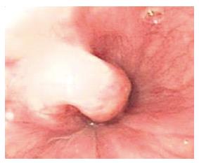
Figure 1 Endoscopic image of esophageal stromal tumors.
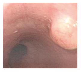
Figure 2 Endoscopic image of leiomyomas.
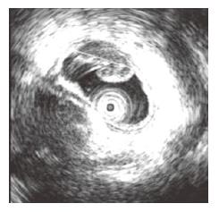
Figure 3 EUS image of esophageal stromal tumors: originated from muscularis propria.
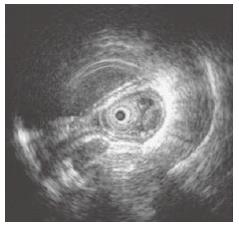
Figure 4 EUS image of leiomyomas: originated from muscularis mucosa.
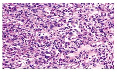
Figure 5 Malignant esophageal stromal tumor: tumor cells were intensely stained, but there was no visible mitoschisis.
(HE × 200).
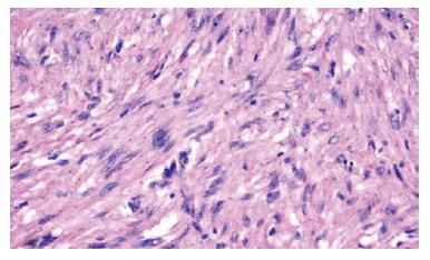
Figure 6 Esophageal leiomyoma: cells were all spindle, with abundant eosinophilic cytoplasm (HE × 200).
Figure 7 Expression of CD117(× 200) and CD 34 (× 400) in malignant esophageal stromal tumor: showing yellow or brown granules in cell cytoplasm and (or) membrane.
A: CD117; B: CD34.
Figure 8 Expression of SMA (A) and Desmin (B) in esophageal leiomyomas (× 200): showing yellow or brown granules in cell cytoplasm.
- Citation: Zhu X, Zhang XQ, Li BM, Xu P, Zhang KH, Chen J. Esophageal mesenchymal tumors: Endoscopy, pathology and immunohistochemistry. World J Gastroenterol 2007; 13(5): 768-773
- URL: https://www.wjgnet.com/1007-9327/full/v13/i5/768.htm
- DOI: https://dx.doi.org/10.3748/wjg.v13.i5.768


