Copyright
©2007 Baishideng Publishing Group Co.
World J Gastroenterol. Feb 7, 2007; 13(5): 683-691
Published online Feb 7, 2007. doi: 10.3748/wjg.v13.i5.683
Published online Feb 7, 2007. doi: 10.3748/wjg.v13.i5.683
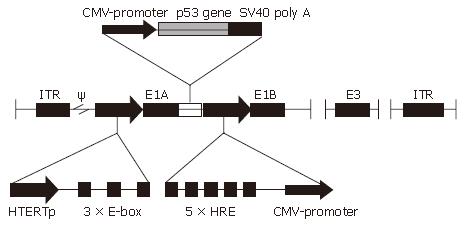
Figure 1 Schematic diagram of the CNHK500-p53 adenoviral construct.
A 310-bp fragment of human telomerase reverse transcriptase (hTERT) promoter with three E-boxes (CACGTG) downstream of the core sequence replaced the endogenous E1A promoter (digested with NotI and XhoI) to control the expression of E1A. A 241-bp fragment of hypoxia response element (HRE) promoter replaced the endogenous E1B promoter to control the expression of E1B. A 1805-bp fragment of transgene expression cassette containing cytomegalovirus (CMV) promoter + p53 + SV40 poly A was inserted into the downstream of E1A (digested with AgeI + NotI) to generate CNHK500-p53. ITR, inverted terminal repeat; ψ, the adenovirus 5 packing signal.
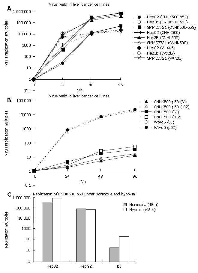
Figure 2 Selective replication of CNHK500-p53 in vitro.
A: Human HCC cell lines HepG2, Hep3B, and SMMC-7721 were infected with CNHK500-p53 at a MOI of 5. Cells and media were harvested, and lysates were prepared from each group at diverse time points 0 h, 24 h, 48 h, and 96 h. Viral titers were measured with the tissue culture infectious dose 50 method, normalized with that at the beginning of infection, and shown as multiples. CNHK500-p53 replicated similarly as CNHK500 and WtAd5 in all of the tested telomerase-positive cancer cells; B: Comparison of replication capability of CNHK500-p53, CNHK500 and WtAd5 in telomerase-negative normal cell lines. At diverse time points 0 h, 24 h, 48 h, and 96 h after infection, cells and medium were harvested, and viral titers were measured as described previously. In all of the tested normal cell lines, the replication capability of CNHK500-p53 and CNHK500 was severely attenuated than that of WtAd5; C: Forty-eight hours after infection with CNHK500-p53 in HepG2, Hep3B and BJ, CNHK500-p53 showed enhanced replication ability both in HepG2, Hep3B and in BJ under hypoxia condition, but was higher in HepG2, Hep3B than in BJ.
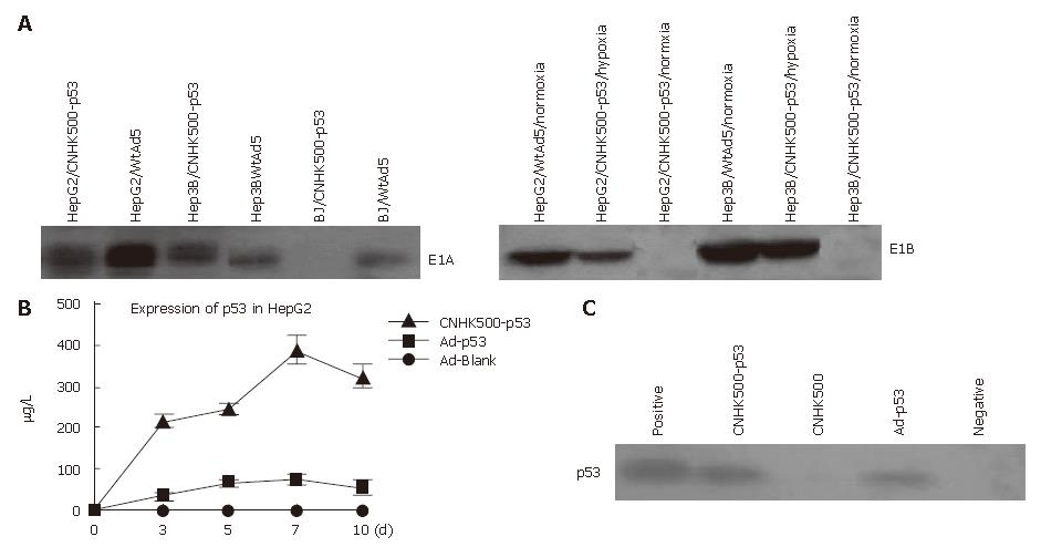
Figure 3 A: E1A and E1B expression identified by Western blot demonstrating that all HCC cells infected with CNHK500-p53 or WtAd5 were positive for E1A expression, however, normal BJ cells were negative for E1A expression when they were infected with CNHK500-p53, and positive only when they were infected with WtAd5, while E1B of CNHK500-p53 was only expressed under hypoxia condition in HepG2 and Hep3B, and expressed both under normal and hypoxia condition with WtAd5; B: ELISA assay showing that p53 protein secreted from a HepG2, infected with CNHK500-p53, was significantly higher than that infected with nonreplicative adenovirus Ad-p53 and Ad-Blank in vitro (P < 0.
05); C: Western blot showing enhanced p53 expression in HepG2 infected with CNHK500-p53.
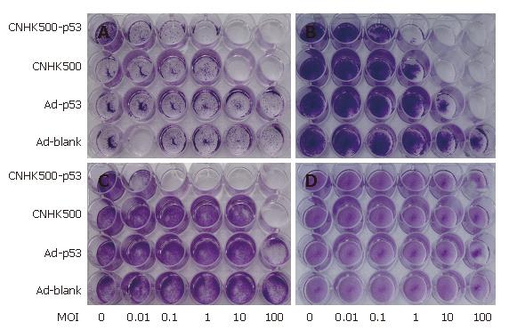
Figure 4 Cytopathic effects associated with CNHK500-p53, CNHK500, Ad-p53, and Ad-Blank infection in HCC cell lines HepG2 (A), Hep3B (B), Bel-7402 (C), and normal cell line BJ (D).
Seven days after virus infection, all the cells were stained with crystal violet and photographed. Comparison with other viruses, CNHK500-p53 showed the strongest selective cytolysis against HCC cell lines. Infection with CNHK500-p53 at MOI of 0.1-1 was sufficient to induce its lytic effects in Bel-7402, HepG2, and Hep3B, although the sensitivity varied among the cell types. In contrast, no apparent cytopathic effects were observed in BJ even at MOI of 100 after CNHK500-p53 infection, which was similar to other viruses.
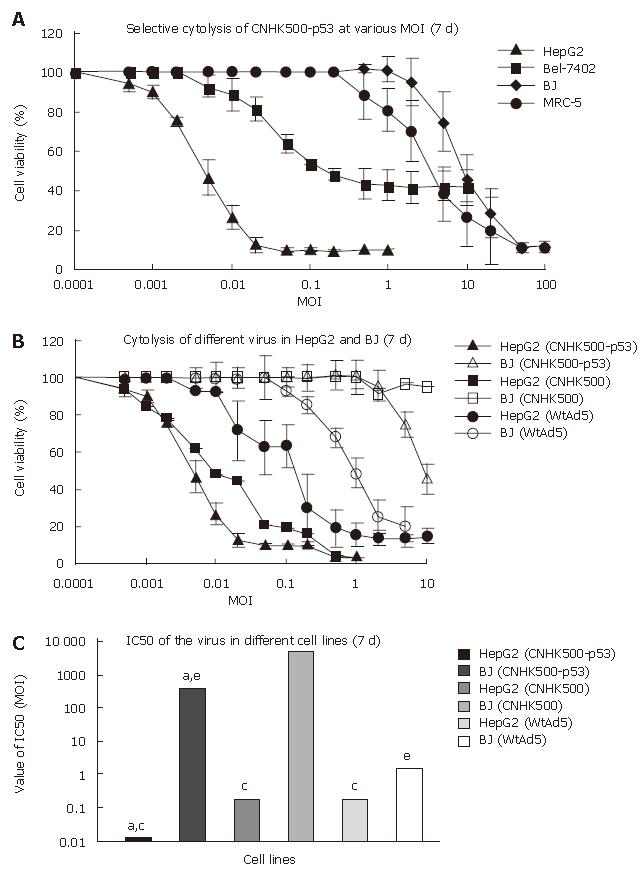
Figure 5 Oncolytic efficacy induced by CNHK500-p53 infection evaluated by MTT assay.
Statistical analysis was performed using Student’s t test for differences among groups. Statistical significance was defined as P < 0.05. A: HCC cell lines HepG2, Bel-7402 and normal cell lines BJ, MRC-5 were infected at different MOI of 0.001-100 pfu/cell of CNHK500-p53. Cell viability was measured by MTT assay 7 d after infection. CNHK500-p53 induced a more powerful oncolysis in HCC cell lines than in normal cell lines (P < 0.05); B: Seven days after infection with virus, CNHK500-p53 not only showed more powerful cytolysis than CNHK500 and WtAd5 in HepG2, but also less profound cytolysis than WtAd5 in BJ (P < 0.05); C: IC50 of CNHK500-p53, CNHk500, and WtAd5 demonstrated significant differences among groups with a,c,eP < 0.05.
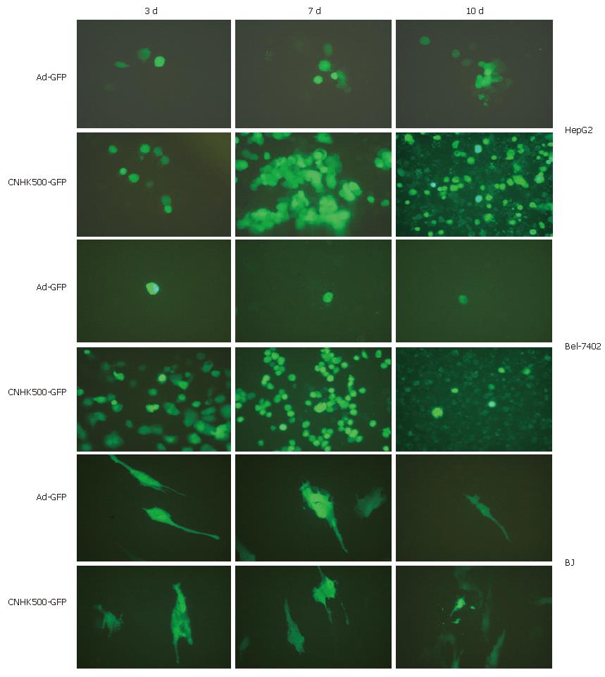
Figure 6 Fluorescent photos of HCC cell lines HepG2, Bel-7402 and normal cell line BJ infected with replicative virus CNHK500-GFP and nonreplicative virus Ad-GFP (200 magnifications).
-
Citation: Zhao HC, Zhang Q, Yang Y, Lu MQ, Li H, Xu C, Chen GH. p53-expressing conditionally replicative adenovirus CNHK500-p53 against hepatocellular carcinoma
in vitro . World J Gastroenterol 2007; 13(5): 683-691 - URL: https://www.wjgnet.com/1007-9327/full/v13/i5/683.htm
- DOI: https://dx.doi.org/10.3748/wjg.v13.i5.683


