Copyright
©2007 Baishideng Publishing Group Co.
World J Gastroenterol. Dec 28, 2007; 13(48): 6598-6602
Published online Dec 28, 2007. doi: 10.3748/wjg.v13.i48.6598
Published online Dec 28, 2007. doi: 10.3748/wjg.v13.i48.6598
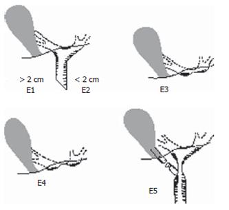
Figure 1 Strasberg classification of bile duct injury.
E1: Transected main bile duct with a stricture more than 2 cm from the hilus; E2: Transected main bile duct with a stricture less than 2 cm from the hilus; E3: Stricture of the hilus with right and left ducts in communication; E4: Stricture of the hilus with separation of right and left ducts; E5: Stricture of the main bile duct and the right posterior sectoral duct.
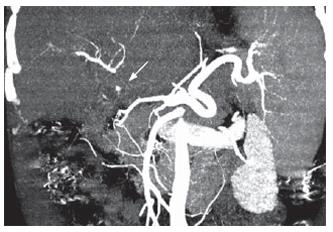
Figure 2 Computed Tomography angiography (CTA) of one type E4 injury patient displayed the lesion of the proper hepatic artery (arrow), although some compensatory collateral arterial blood supply from the left gastric artery could be identified.
This patient received liver transplantation.
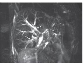
Figure 3 MRCP appearance of one type E3 injury patient showing dilation of intrahepatic bile ducts (arrows).
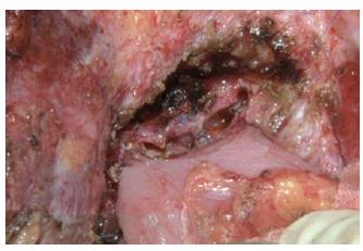
Figure 4 Plastic reconstruction in one type E3 patient merging several bile duct openings into one at the hilum after removal of the scarred ducts (the same patient in Figure 3).
- Citation: Yan JQ, Peng CH, Ding JZ, Yang WP, Zhou GW, Chen YJ, Tao ZY, Li HW. Surgical management in biliary restricture after Roux-en-Y hepaticojejunostomy for bile duct injury. World J Gastroenterol 2007; 13(48): 6598-6602
- URL: https://www.wjgnet.com/1007-9327/full/v13/i48/6598.htm
- DOI: https://dx.doi.org/10.3748/wjg.v13.i48.6598


