Copyright
©2007 Baishideng Publishing Group Inc.
World J Gastroenterol. Dec 21, 2007; 13(47): 6404-6409
Published online Dec 21, 2007. doi: 10.3748/wjg.v13.i47.6404
Published online Dec 21, 2007. doi: 10.3748/wjg.v13.i47.6404
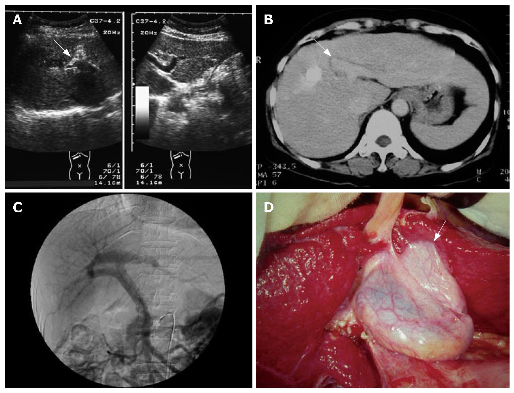
Figure 1 A 50-year-old woman with chronic hepatitis C.
Right trise-gmentectomy and cholecy-stectomy were performed due to the right lobe liver tumor. A: US demonstrated a club-shaped portal vein (arrow) over the right lobe of the liver and no umbilical portion over the left lobe of the liver; B: Enhanced CT demonstrated enlarged left lateral segment and a club-shaped portal vein (arrow) over the right side of the liver; C: Angiography demonstrated trifurcation type of the portal vein; D: Intraoperative photography demo-nstrated gallbladder over left side of ligamentum teres and adhesions on the left lateral segment (arrow).
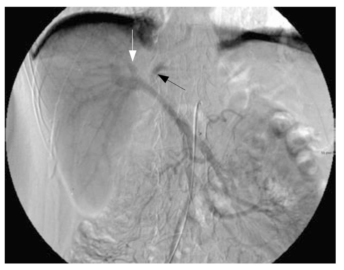
Figure 2 A 55-year-old man with HCC, chronic hepatitis B and C.
Angio-portography demonstrated bifurcation with a small branch of the left portal vein (black arrow), segmental IV branch of the portal vein (white arrow), and a club-shaped form of the right portal vein with many branches.
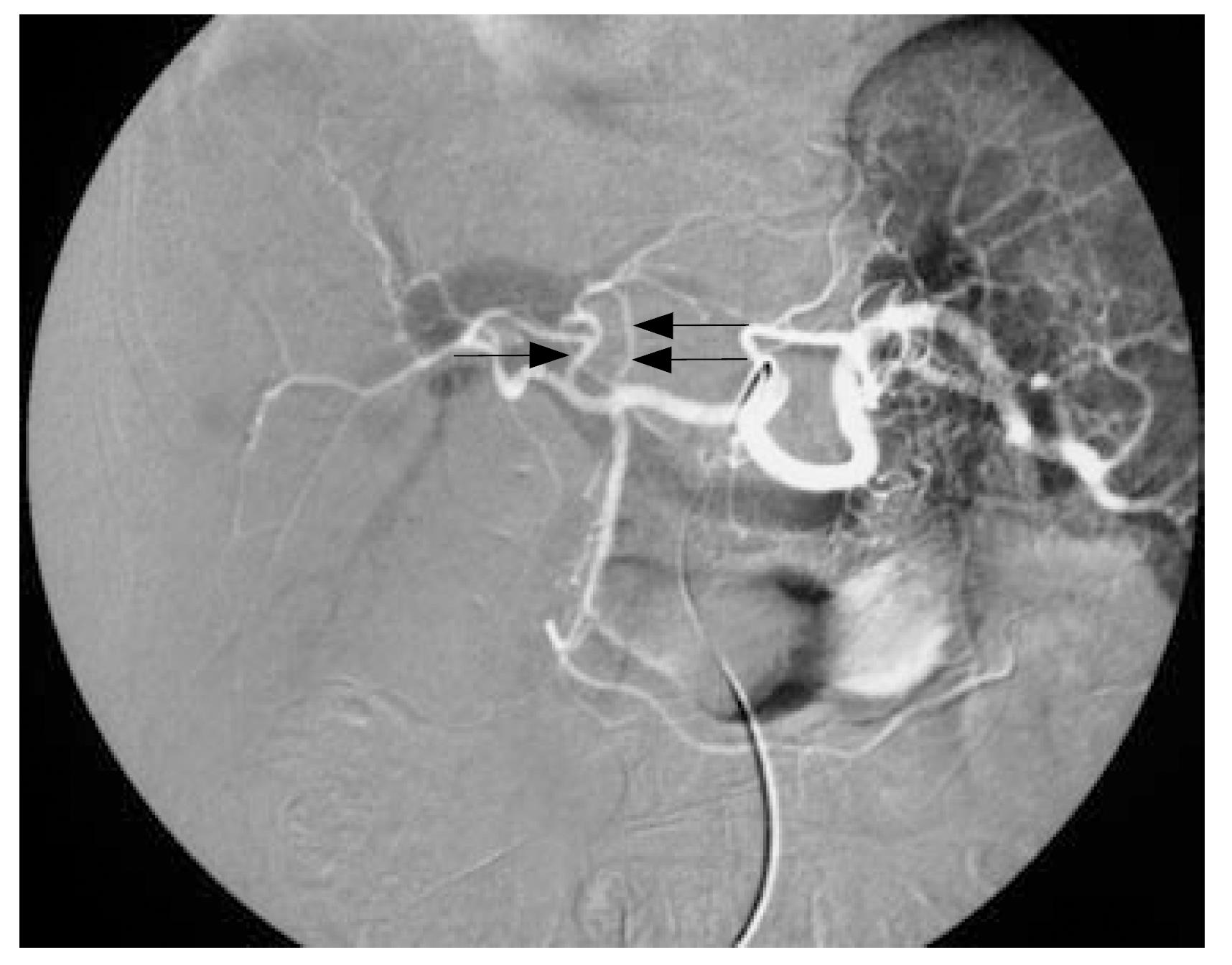
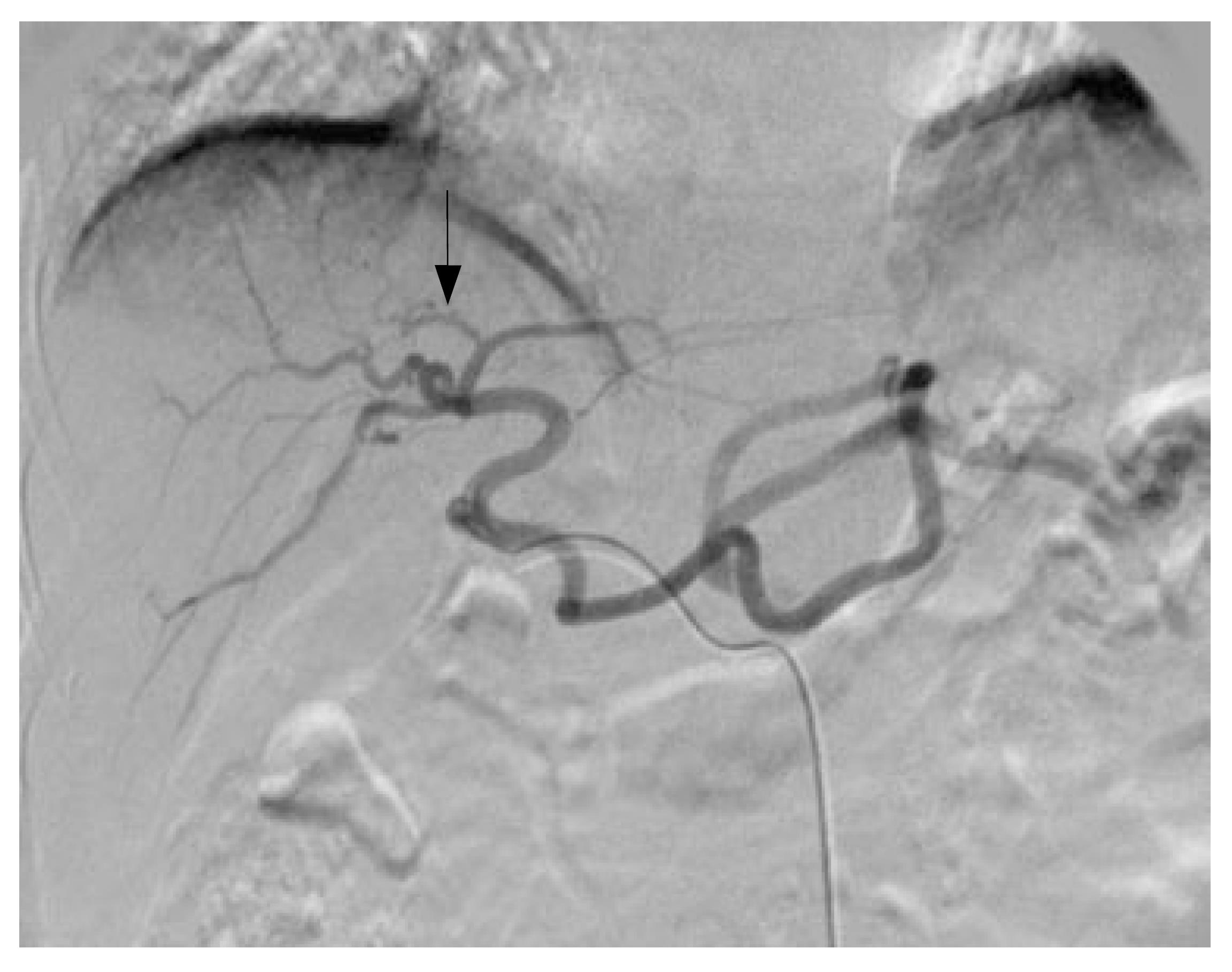
Figure 3 A 59-year-old woman with HCC in the segment VI of the liver and chronic hepatitis C.
Angio-portography with substrate on arterial phase. The hepatic arteries (white vessels) followed the portal veins (black vessels). The segmental IV artery (arrow) was demonstrated. Right anterior side liver parenchyma was supplied from the segmental IV artery. The left hepatic artery (double arrows) originated from the common hepatic artery near the orifice of the gastroduodenal artery.
Figure 4 Maetini et al have described three types of portal vein anomalies with the umbilical portion supplying the right anterior portion.
In type A, the umbilical portion is over the right side of the Cantlie line. In type B, the umbilical portion lies on the Cantlie line. In type C, the umbilical portion arises from the left portal vein with anterior segmental branches. As the ligamentum teres moves from the type C to the type A, the gallbladder moves to the left of the ligamentum teres.
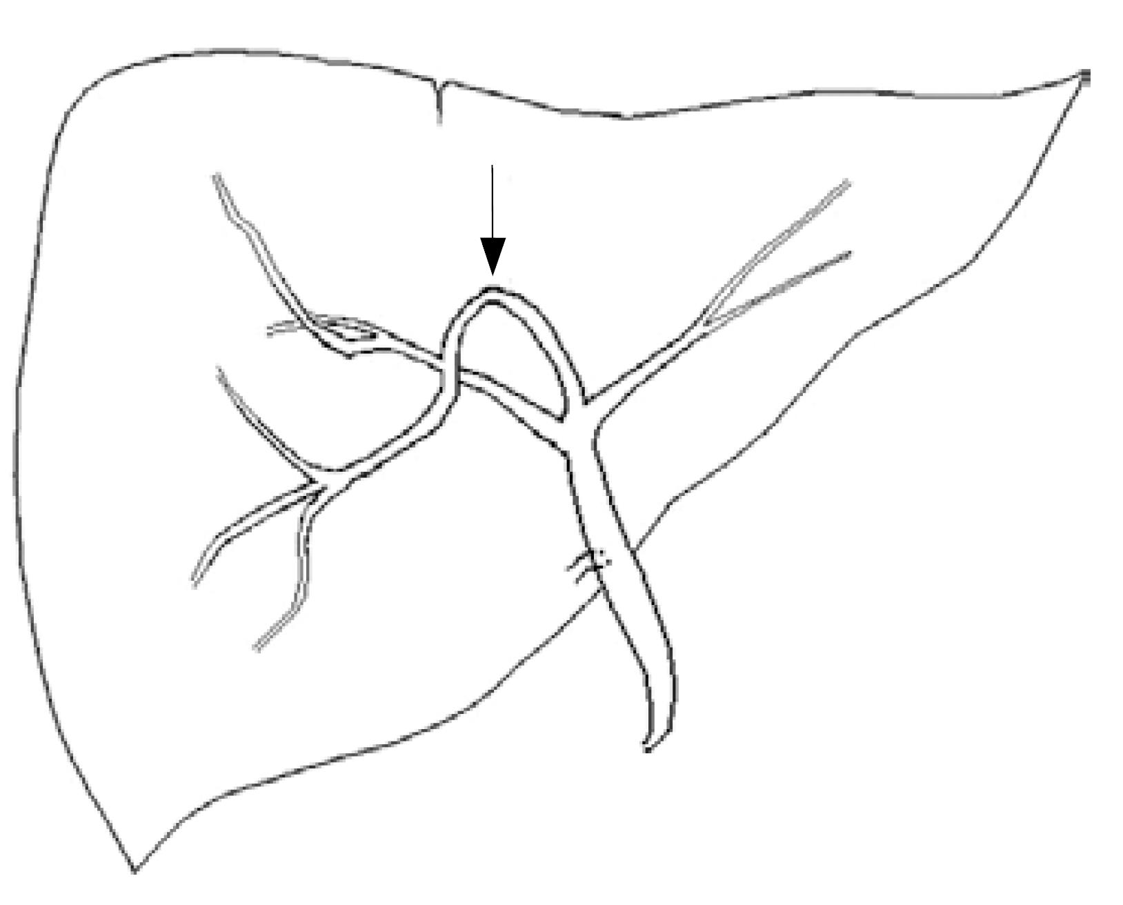
Figure 5 The right anterior segmental bile duct ran downward and ventrally formed a reverse U-shape (arrow), which followed the right anterior portal vein and lay in the groove for the ligamentum teres in left-sided gallbladder.
Maetani et al believe that the gallbladder on the left side of the ligamentum teres is simply because the latter deviated to the right.
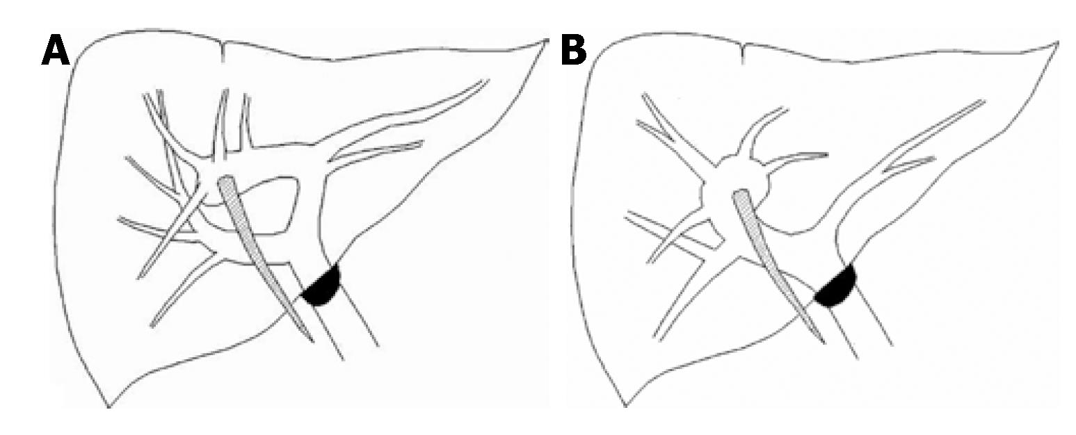
Figure 6 A: The trifurcation type of portal vein anomaly in a left-sided gallbladder; B: Bifurcation type of portal vein anomaly in a left-sided gallbladder.
The abnormal intrahepatic portal venous branches could not be determined into these two types was divided into the other type.
Figure 7 A 64-year-old man with HCC.
The segmental IV artery (arrow) from the left hepatic artery is demonstrated.
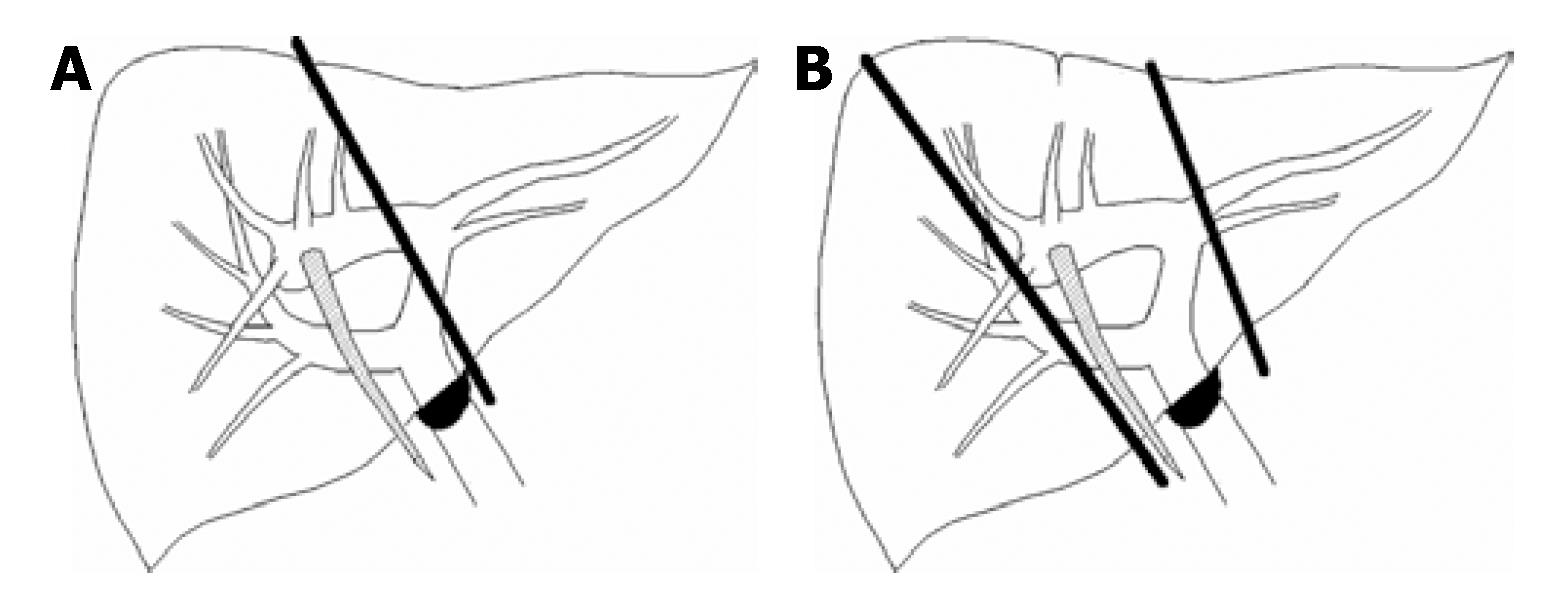
Figure 8 Trifurcation type of portal vein anomaly.
A: If left hepatectomy is performed, the portal veins draining three-quarters of the liver parenchyma will be ligated. Hepatic necrosis may occur over right anterior portion of the liver parenchyma and contribute to hepatic failure; B: Left lateral segmentectomy or right hepatectomy is safe in this type portal vein anomaly.
- Citation: Hsu SL, Chen TY, Huang TL, Sun CK, Concejero AM, Tsang LLC, Cheng YF. Left-sided gallbladder: Its clinical significance and imaging presentations. World J Gastroenterol 2007; 13(47): 6404-6409
- URL: https://www.wjgnet.com/1007-9327/full/v13/i47/6404.htm
- DOI: https://dx.doi.org/10.3748/wjg.v13.i47.6404


