Copyright
©2007 Baishideng Publishing Group Inc.
World J Gastroenterol. Dec 21, 2007; 13(47): 6327-6332
Published online Dec 21, 2007. doi: 10.3748/wjg.v13.i47.6327
Published online Dec 21, 2007. doi: 10.3748/wjg.v13.i47.6327
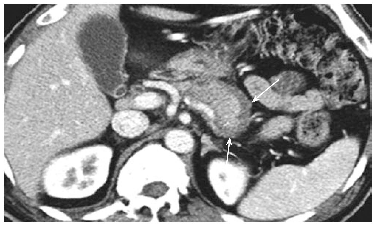
Figure 1 Contrast enhanced CT showing sausage-like swelling of the pancreatic tail along with a surrounding low attenuation rim (arrows).
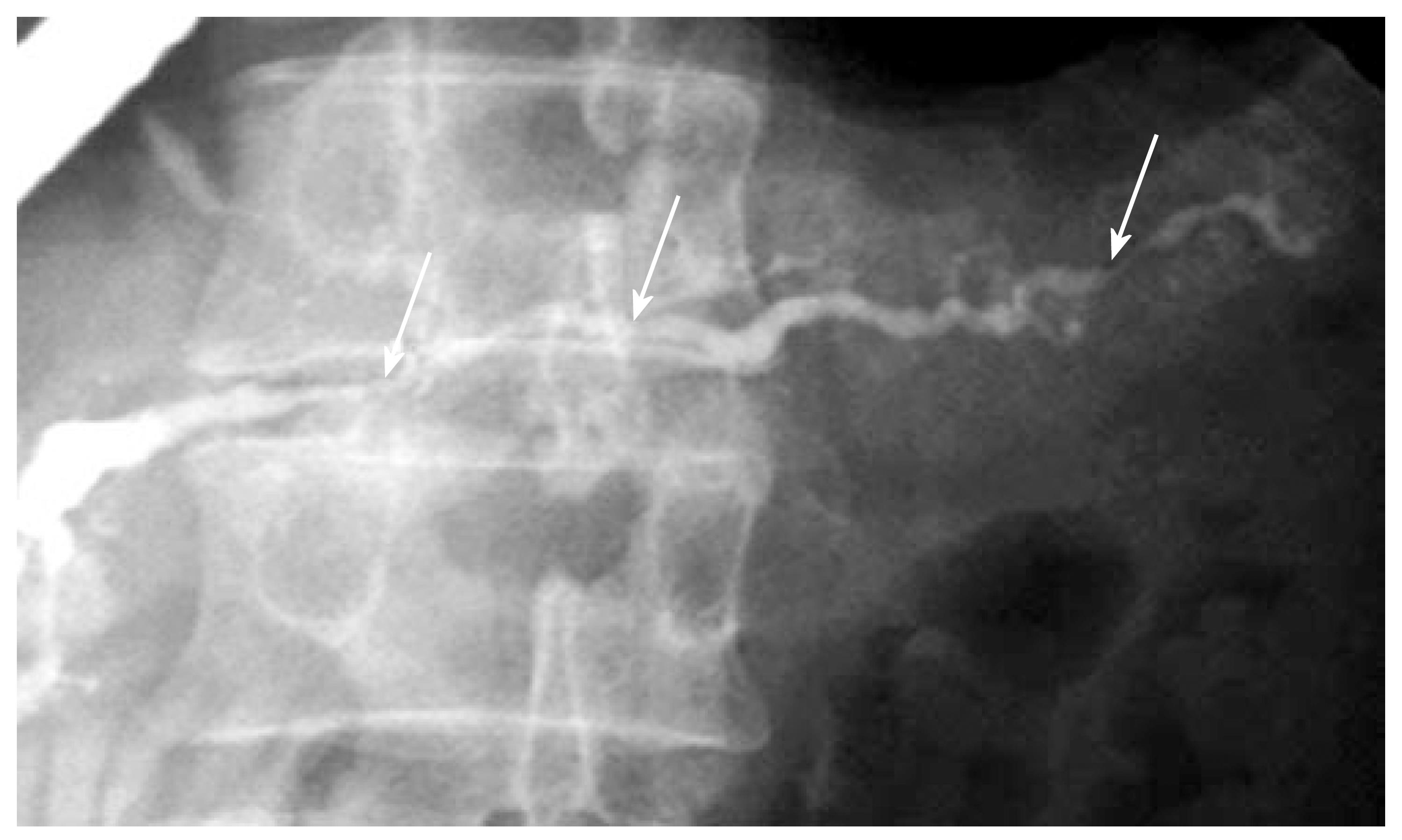
Figure 2 Direct pancreatography delineating a pancreatic duct with multiple segments of narrowing (arrows).
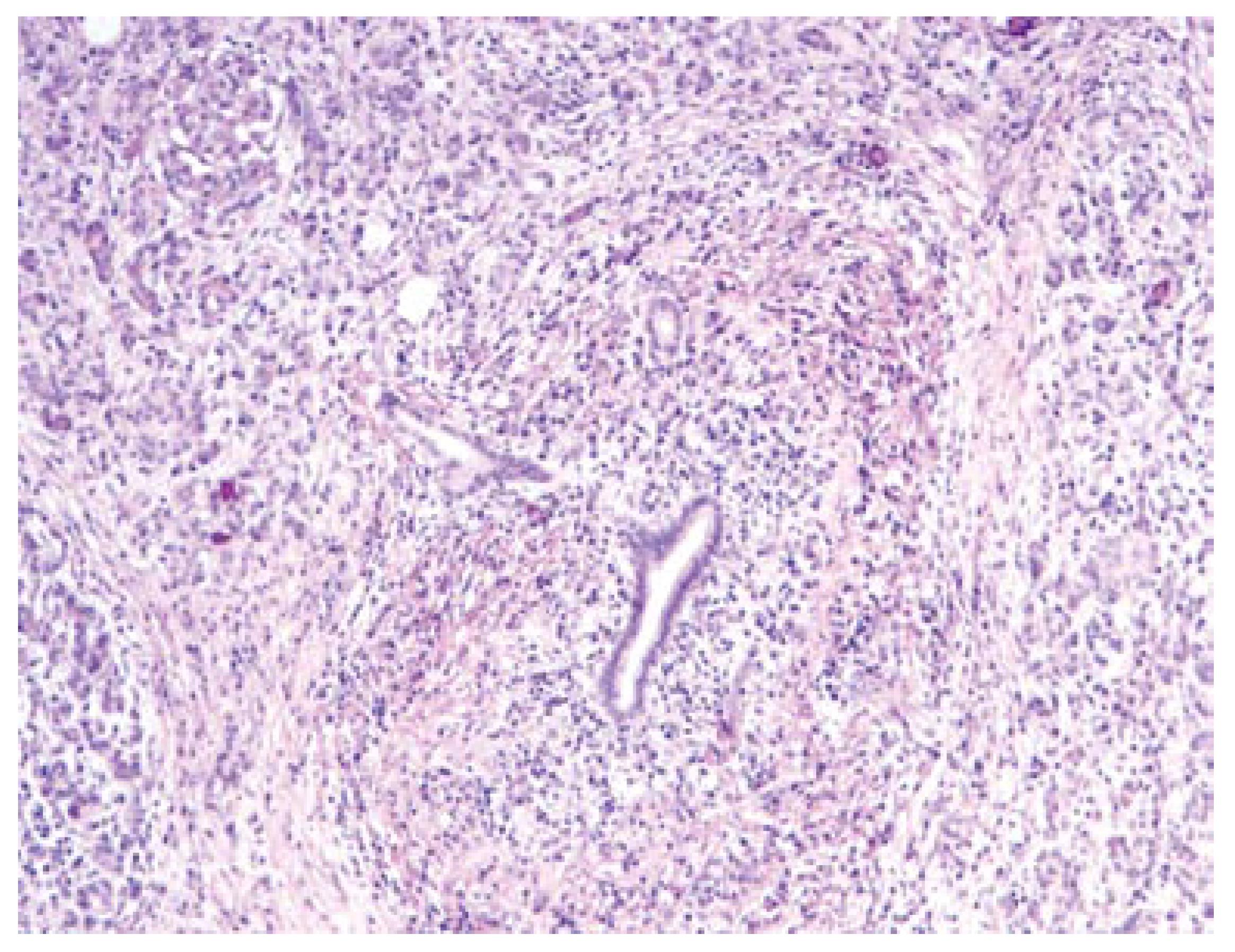
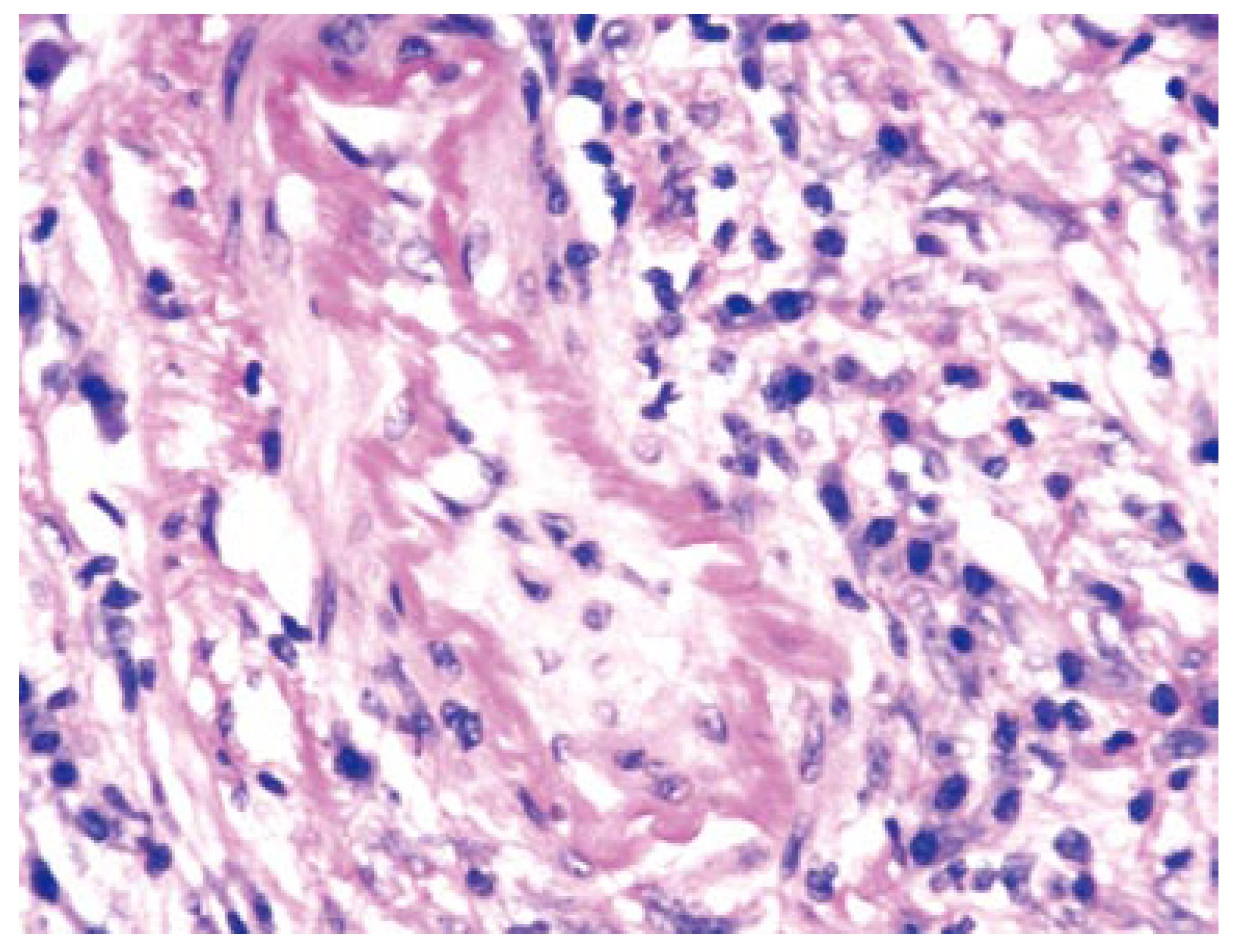
Figure 3 Diffuse pancreatic lymphoplasmacytic infiltrate with early fibrosis and atrophy.
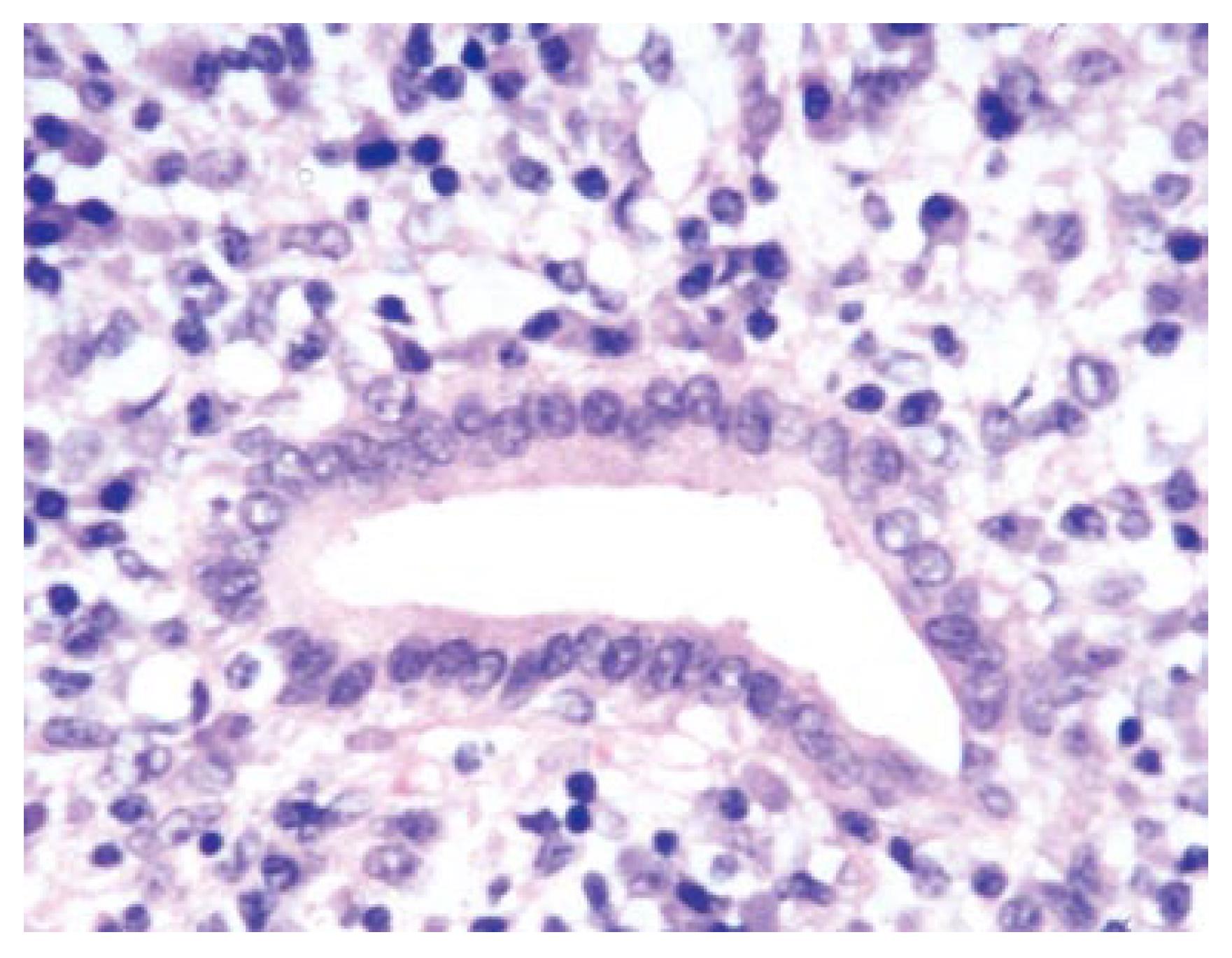
Figure 4 Small pancreatic duct surrounded by a cuff of lymphocytes and plasma cells that is extending into the atrophic pancreatic parenchyma.
Figure 5 Medium sized artery with infiltration of the vessel wall by lymphocytes and plasma cells.
- Citation: Zandieh I, Byrne MF. Autoimmune pancreatitis: A review. World J Gastroenterol 2007; 13(47): 6327-6332
- URL: https://www.wjgnet.com/1007-9327/full/v13/i47/6327.htm
- DOI: https://dx.doi.org/10.3748/wjg.v13.i47.6327


