Copyright
©2007 Baishideng Publishing Group Co.
World J Gastroenterol. Nov 28, 2007; 13(44): 5805-5812
Published online Nov 28, 2007. doi: 10.3748/wjg.v13.i44.5805
Published online Nov 28, 2007. doi: 10.3748/wjg.v13.i44.5805
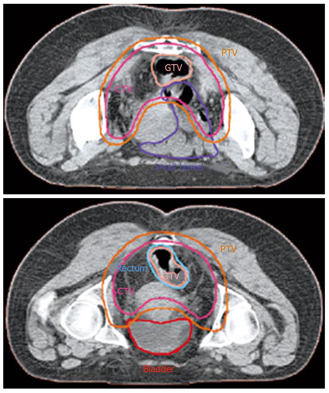
Figure 1 The GTV, PTV and organ at risk (small bowel and bladder) countered on the axial CT slices.
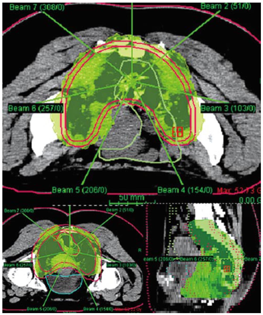
Figure 2 Axial and sagital CT scan images with dose distributions.
The 45 Gy isodose surface (green) encompass the GTV and PTV.
- Citation: Diaz-Gonzalez JA, Arbea L, Aristu J. Rectal cancer treatment: Improving the picture. World J Gastroenterol 2007; 13(44): 5805-5812
- URL: https://www.wjgnet.com/1007-9327/full/v13/i44/5805.htm
- DOI: https://dx.doi.org/10.3748/wjg.v13.i44.5805


