Copyright
©2007 Baishideng Publishing Group Inc.
World J Gastroenterol. Oct 28, 2007; 13(40): 5403-5407
Published online Oct 28, 2007. doi: 10.3748/wjg.v13.i40.5403
Published online Oct 28, 2007. doi: 10.3748/wjg.v13.i40.5403
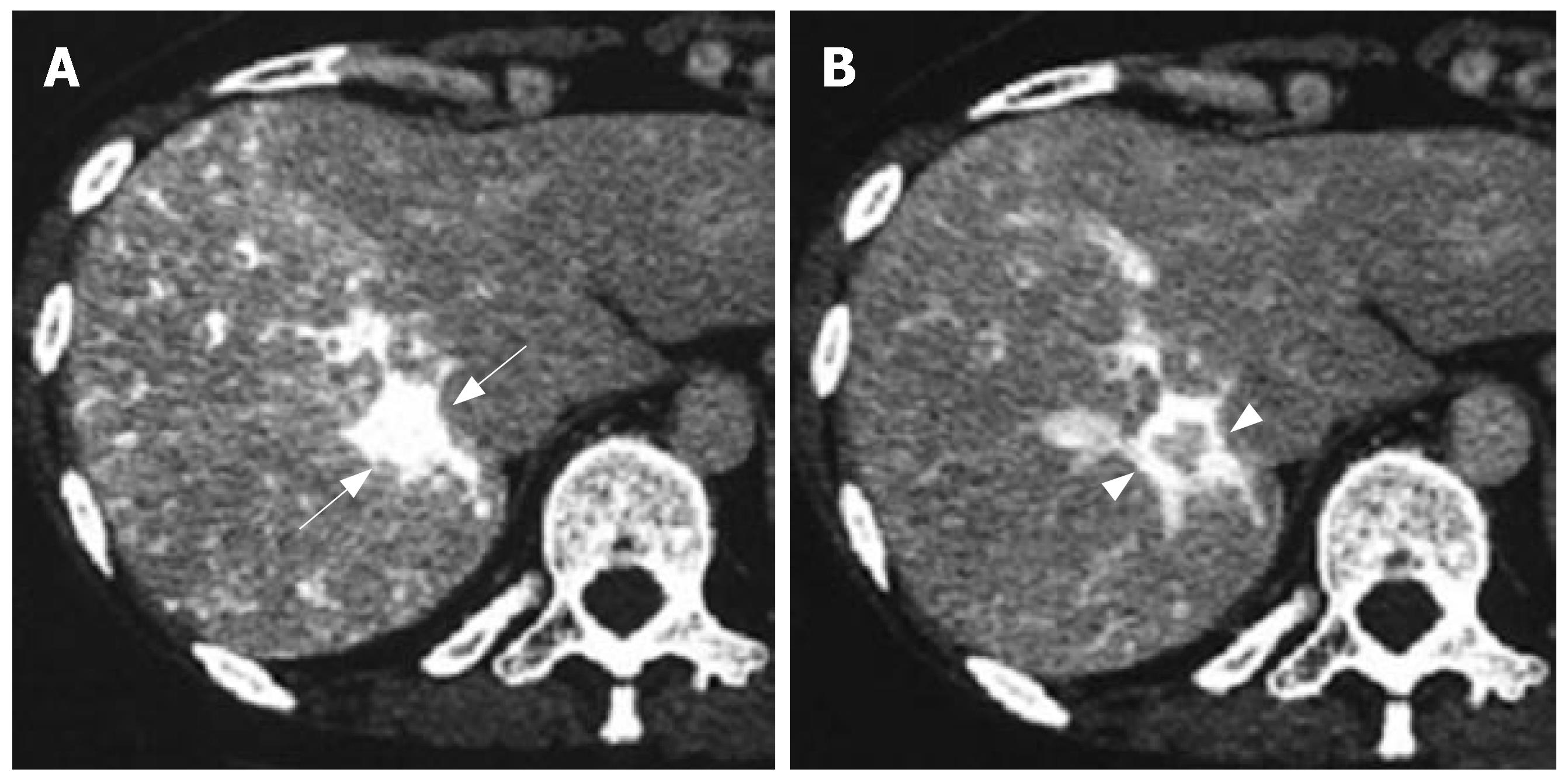
Figure 1 CT angiography showing the conspicuously enhanced lesion in segment 7 through the early phase (arrows) (A) and intensified tumor rim enhancement through the delayed phase along with radial enhancement (arrowheads) (B).
Figure 2 MRI displaying a hypointense signal on T1-weighted imaging (arrow) (A), a hyperintense signal on T2-weighted imaging of the lesion (arrow) (B), and enhanced tumor rim in the delayed phase of MRI (arrow) (C).
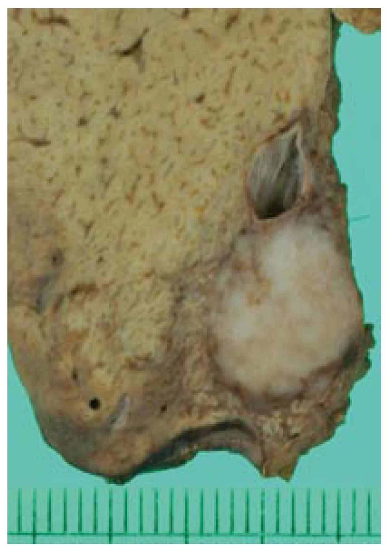
Figure 3 Macroscopy of tumor in segment 7 demonstrating the lesion as a well-circumscribed, white-yellowish nodule on the cut surface.
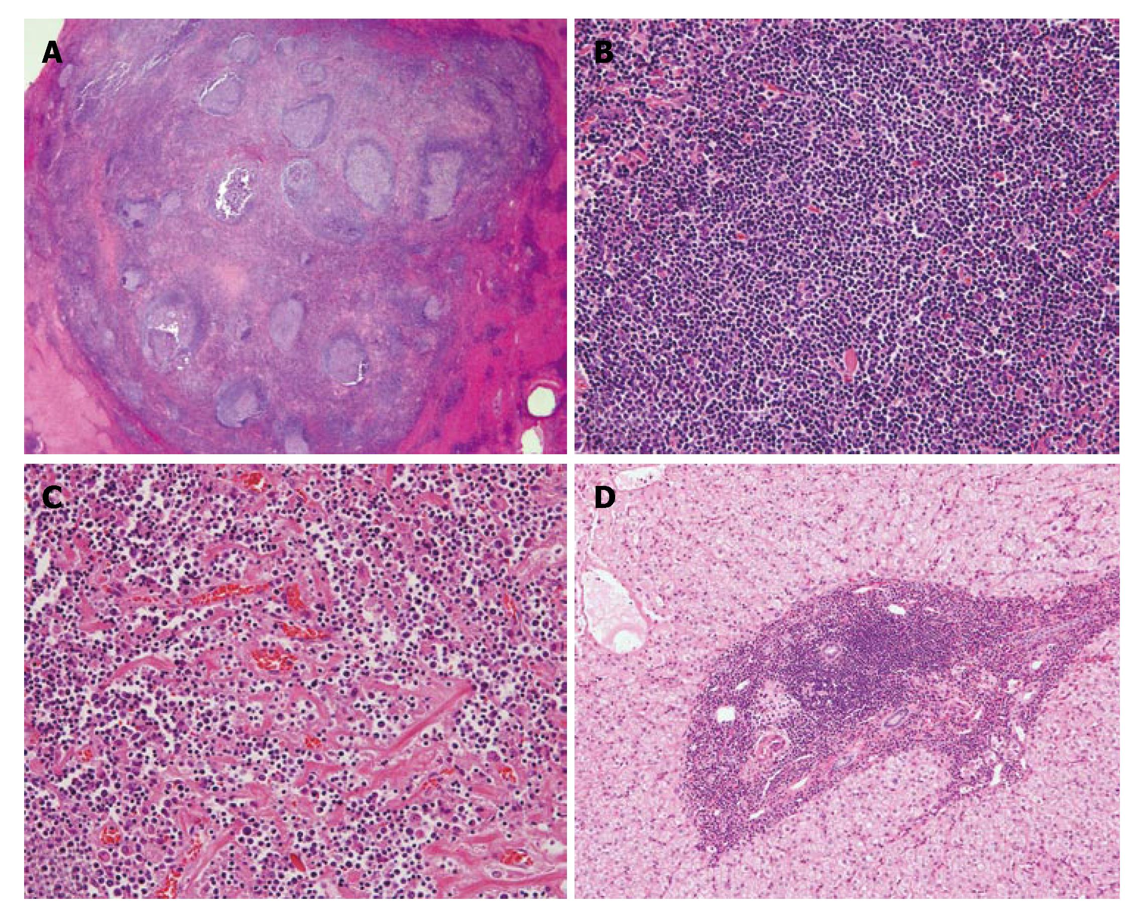
Figure 4 Microscopy revealing lesions comprising massive lymphoid cell infiltration with prominent follicles and hyalinized interfollicular spaces at low magnification (HE, X 2.
5) (A), interfollicular areas mainly comprising mature lymphocytes with no nuclear atypia (HE, X 50) (B), strands of amorphous hyalinized material accompanying capillaries in interfollicular areas (HE, X 50) (C), and portal areas apart from the nodules with irregular expansion of infiltration of small mature lymphocytes (HE, X 25) (D).
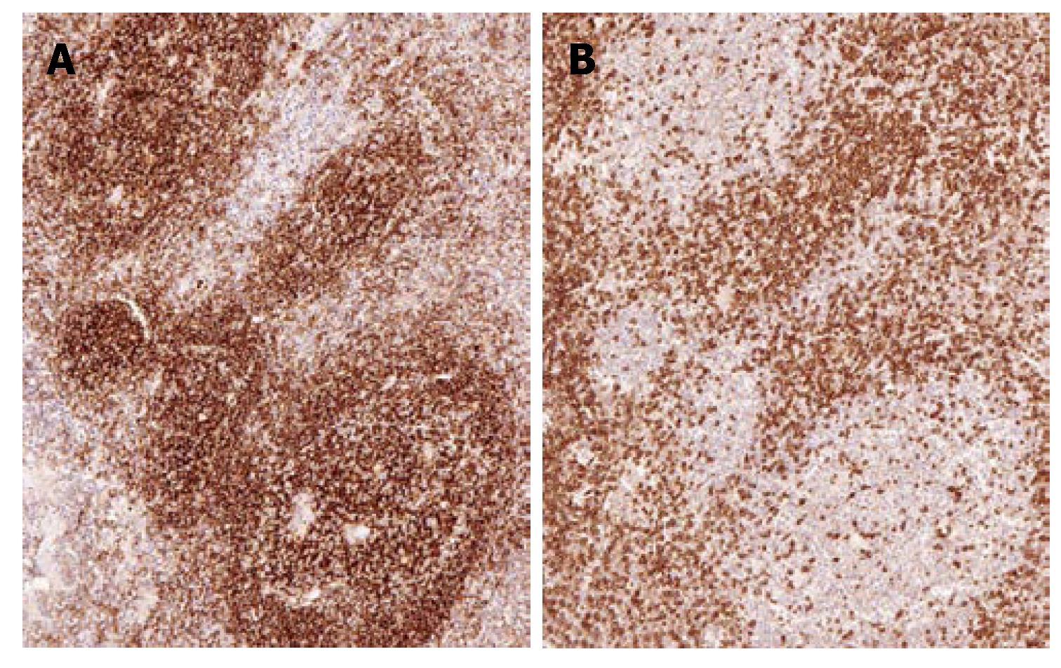
Figure 5 Immunohistochemistry discovering follicles mainly comprising CD20-positive lymphocytes (X 50) (A), and interfollicularly and circumferentially distributed CD3-positive cells compared to follicles (X 50) (B).
- Citation: Machida T, Takahashi T, Itoh T, Hirayama M, Morita T, Horita S. Reactive lymphoid hyperplasia of the liver: A case report and review of literature. World J Gastroenterol 2007; 13(40): 5403-5407
- URL: https://www.wjgnet.com/1007-9327/full/v13/i40/5403.htm
- DOI: https://dx.doi.org/10.3748/wjg.v13.i40.5403


