Copyright
©2007 Baishideng Publishing Group Inc.
World J Gastroenterol. Oct 28, 2007; 13(40): 5384-5390
Published online Oct 28, 2007. doi: 10.3748/wjg.v13.i40.5384
Published online Oct 28, 2007. doi: 10.3748/wjg.v13.i40.5384
Figure 1 A: No evidence of collagen deposition was observed in the liver of the normal group (HE, × 400); B: The collagens deposited in portal areas of liver cirrhosis group (HE, × 400); C: Severe fibrosis with regenerating nodules was observed in liver cirrhosis group (Masson, × 100).
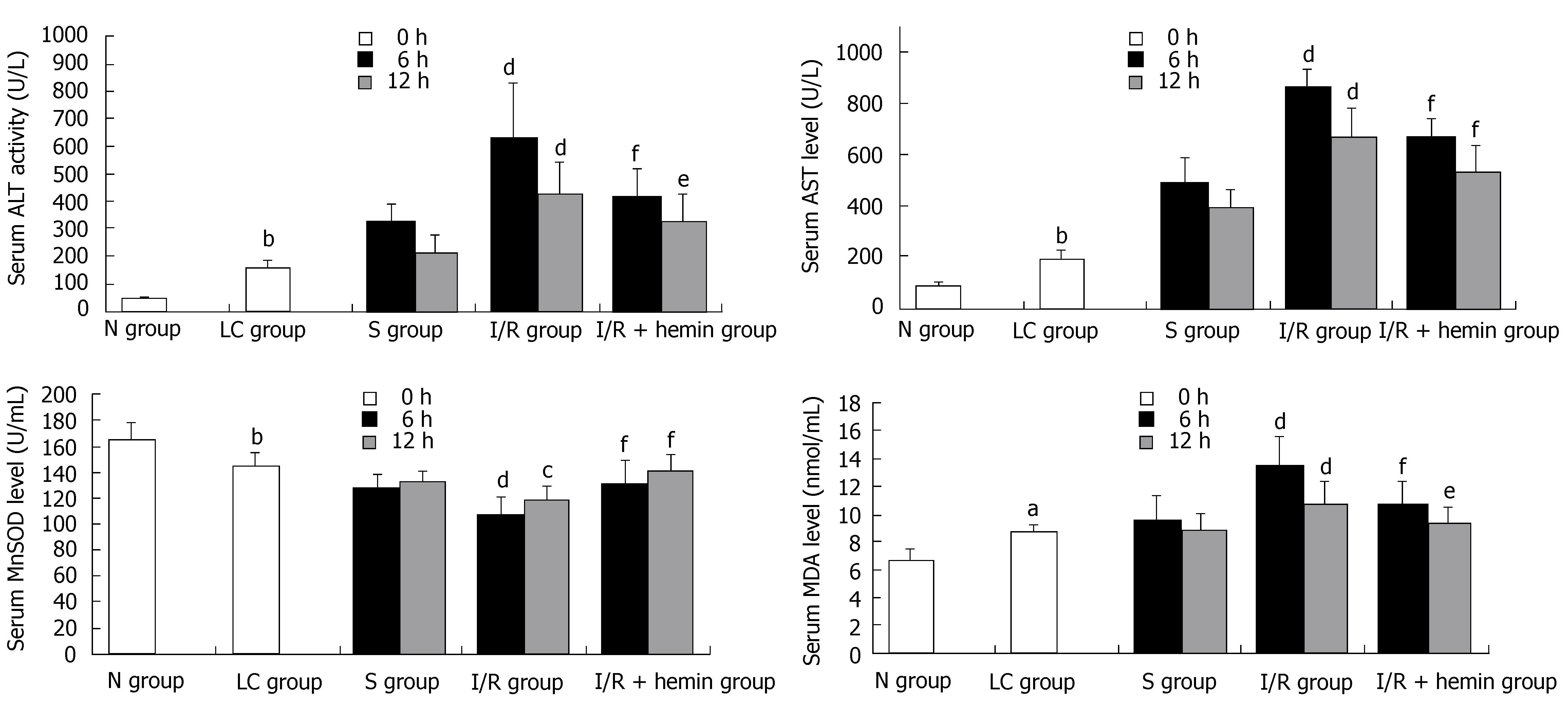
Figure 2 Serum ALT, AST activity, MnSOD and MDA levels of each group.
N group: normal group; LC group: liver cirrhosis group; S group: sham group; I/R group: the rats underwent hepatic ischemia/reperfusion operation; I/R + hemin group: the rats were administered with 30 μmol/kg hemin before hepatic ischemia/reperfusion operation. 6 h: 6 h after reperfusion. 12 h: 12 h after reperfusion. After hepatic ischemia/reperfusion, the ALT, AST and MDA levels were increased and the levels were all decreased after hemin administration. The level of MnSOD was low in I/R group, and after giving hemin, its level in I/R + hemin group was higher than I/R group. Values are mean ± SD. bP < 0.01 vs N group, aP < 0.05 vs N group, dP < 0.01 vs S group, cP < 0.05 vs S group, fP < 0.01 vs I/R group, eP < 0.05 vs I/R group.
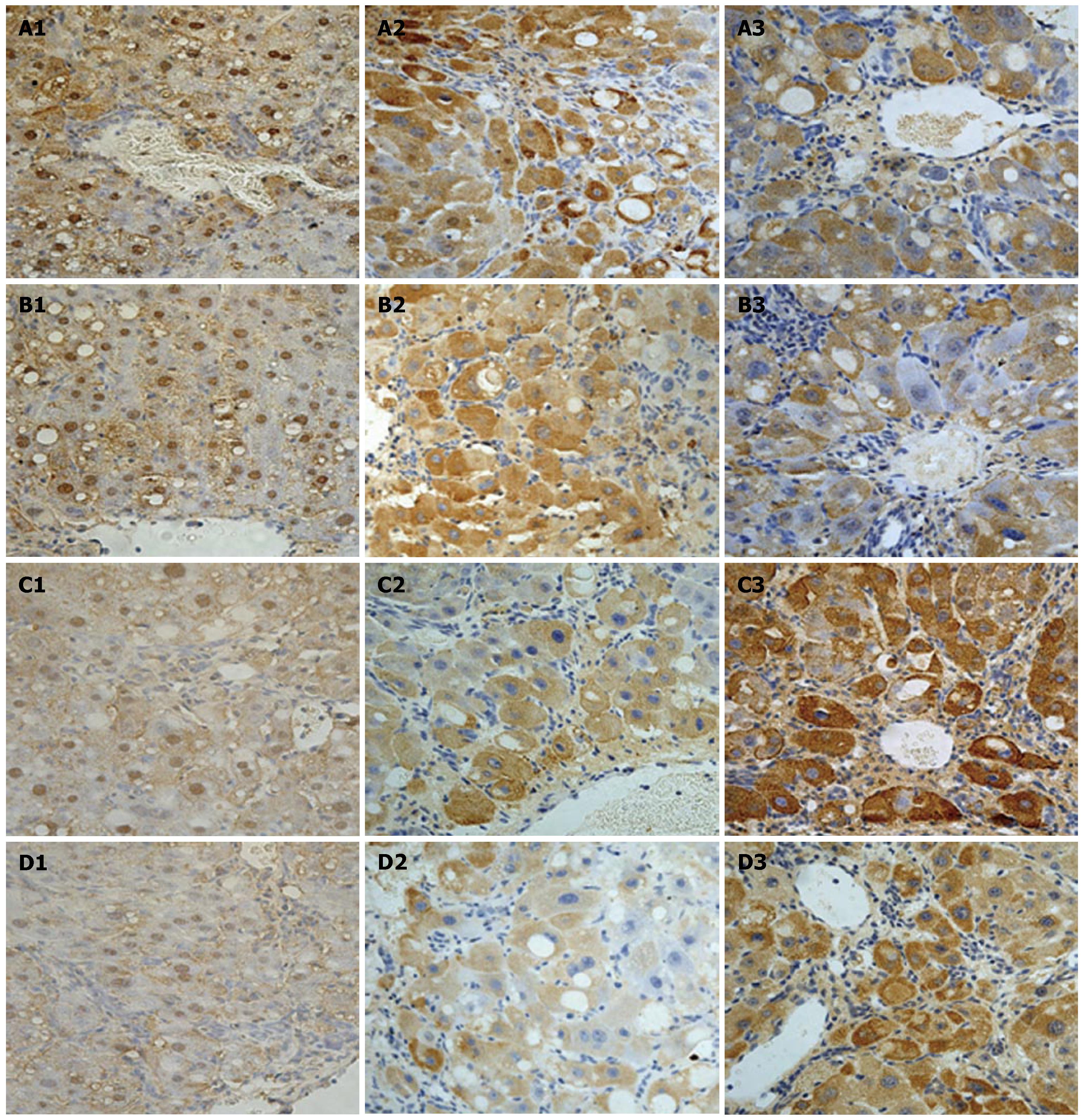
Figure 3 Immunohistochemistry of NF-κB, caspase-3 and HO-1 at 6 h and 12 h after hepatic ischemia/reperfusion in cirrhotic rats.
A: I/R group, 6 h after reperfusion; B: I/R group, 12 h after reperfusion; C: I/R + hemin group, 6 h after reperfusion; D: I/R + hemin group, 12 h after reperfusion; 1: NF-κB: Most positive NF-κB expression was in the nuclear of liver cells. After giving hemin to induce HO-1, the positive expression of NF-κB in I/R + hemin group was decreased compared with I/R group at 6 h after reperfusion (P < 0.01). There is no difference between I/R group and I/R + hemin group at 12 h after reperfusion (P > 0.05) (Figure A1-D1); 2: Caspase-3: The positive caspase-3 expression was in the cytoplasm of hepatic cells. After hemin administration, the caspase-3 expression was decreased at 6 h and 12 h after reperfusion when compared with I/R group (P < 0.01) (Figure A2-D2); 3: HO-1: The positive HO-1 expression was in the plasma of hepatic cells. After hepatic ischemia/reperfusion, the expression of HO-1 was increased in I/R group, and through giving hemin, the expression of HO-1 was higher than I/R group at 6 h and 12 h after reperfusion (P < 0.01) (Figure A3-D3).
- Citation: Xue H, Guo H, Li YC, Hao ZM. Heme oxygenase-1 induction by hemin protects liver cells from ischemia/reperfusion injury in cirrhotic rats. World J Gastroenterol 2007; 13(40): 5384-5390
- URL: https://www.wjgnet.com/1007-9327/full/v13/i40/5384.htm
- DOI: https://dx.doi.org/10.3748/wjg.v13.i40.5384


