Copyright
©2007 Baishideng Publishing Group Inc.
World J Gastroenterol. Oct 28, 2007; 13(40): 5306-5311
Published online Oct 28, 2007. doi: 10.3748/wjg.v13.i40.5306
Published online Oct 28, 2007. doi: 10.3748/wjg.v13.i40.5306
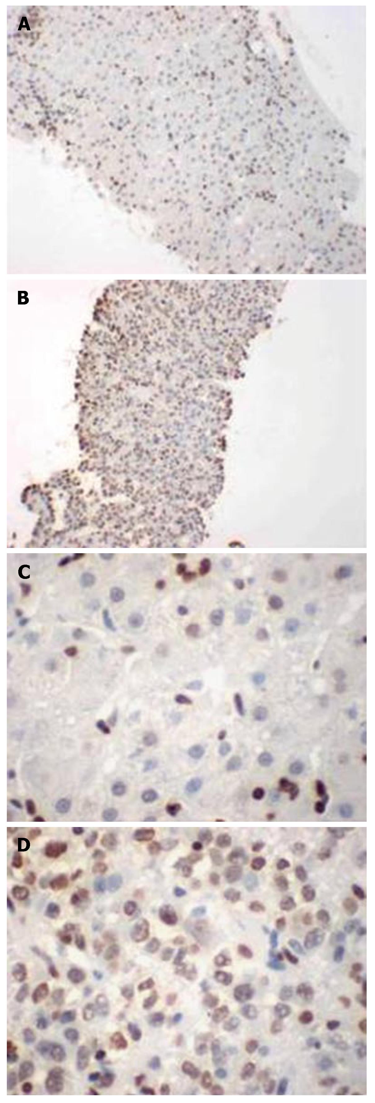
Figure 1 Immunopathological staining of survivin in HCC and peritumoral biopsy tissues.
A: Non-tumor biopsy (х 100); B: Tumor biopsy (х 100); C: Non-tumor biopsy (х 400); D: Tumor biopsy (х 400).
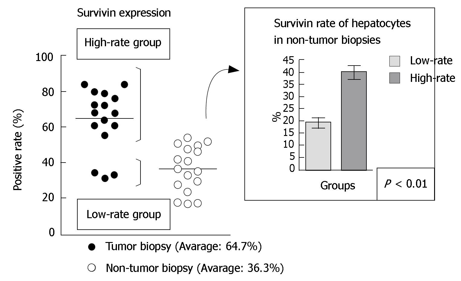
Figure 2 Nuclear survivin expression rates in HCC biopsies and non-tumor biopsies samples.
In tumor biopsies, > 500 survivin-expressing HCC cells were counted in three areas at 100 х magnification using the nuclear labeling index. In non-tumor biopsies, > 500 survivin-expressing hepatocyte cell were counted in three areas at 100 х magnification using the nuclear labeling index.
Figure 3 Expression of survivin mRNA in a hypoxic and anti-cancer drug-containing medium.
The survivin mRNA expression increased under hypoxia, by anti-cancer drug treatment, and in presence of the both conditions in the 6-h culture. The survivin mRNA expression increased under hypoxia and in the combined conditions of hypoxia and anti-cancer drug in the 96-h culture.
Figure 4 Western blotting showing the expression of survivin protein in the combined conditions of hypoxia and anti-cancer drugs in the 96-h culture.
Survivin expression increased under anti-cancer drug-containing medium. Moreover, survivin further increased after the administration of a combination of hypoxia and anti-cancer drug. A: Normoxia; B: Hypoxia; C: Normoxia + 0.1 μmol/L EPI; D: Hypoxia + 0.1 μmol/L EPI.
- Citation: Mamori S, Asakura T, Ohkawa K, Tajiri H. Survivin expression in early hepatocellular carcinoma and post-treatment with anti-cancer drug under hypoxic culture condition. World J Gastroenterol 2007; 13(40): 5306-5311
- URL: https://www.wjgnet.com/1007-9327/full/v13/i40/5306.htm
- DOI: https://dx.doi.org/10.3748/wjg.v13.i40.5306


