Copyright
©2007 Baishideng Publishing Group Inc.
World J Gastroenterol. Oct 21, 2007; 13(39): 5261-5266
Published online Oct 21, 2007. doi: 10.3748/wjg.v13.i39.5261
Published online Oct 21, 2007. doi: 10.3748/wjg.v13.i39.5261
Figure 1 RT-PCR analysis of periostin mRNA expression in human gastric cancer (T) and normal gastric tissues (N).
Periostin was differentially expressed in gastric cancer tissues (T) compared with normal gastric tissues (N) from the same patient. The expression of β-actin was used as an internal control.
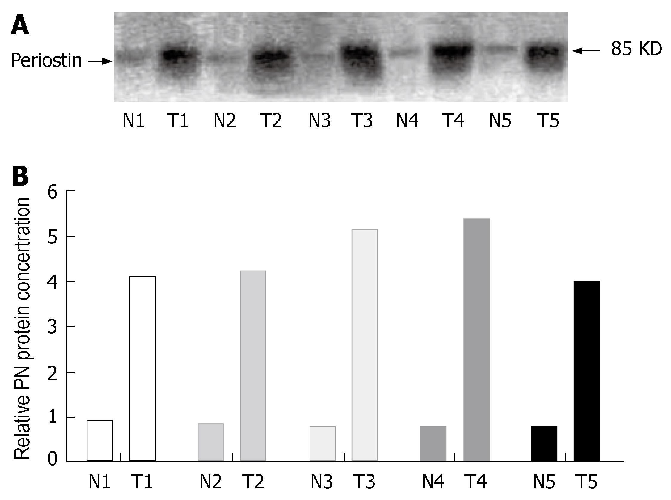
Figure 2 Periostin protein expression in normal and cancer tissue samples.
Tissue extracts from normal or gastric cancer tissue samples were subjected to immunoblot analysis with a polyclonal antiperiostin antibody. The results shown in the T and N lanes are for cancer and normal tissues, respectively, from which RT-PCR was performed (A). Over fivefold more periostin was detected in gastric cancer tissues than in normal tissues from the same patient (B).
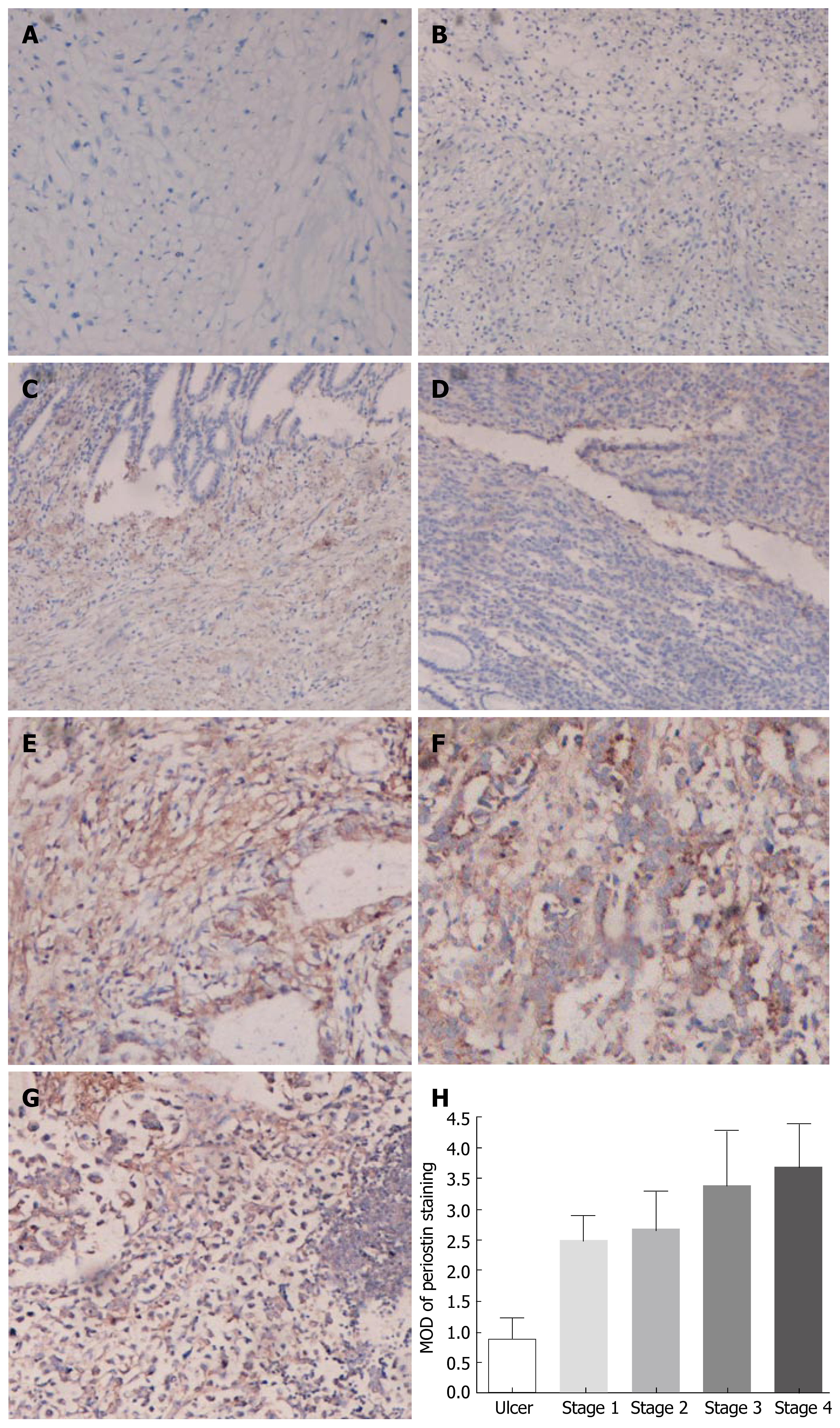
Figure 3 Immunohistochemical analysis of periostin expression in normal gastric tissues, benign gastric ulcers, gastric cancer tissues, as well as lymphoid metastasis from gastric cancer.
The tissue sections were immunostained with a polyclonal antibody. The positive staining for periostin protein is shown with a brown color. All sections were counterstained with hematoxylin showing a blue color. (A) Negative control; (B) benign gastric ulcer; (C) stageIgastric cancer; (D) stageIIgastric cancer; (E) stage III gastric cancer; (F) stage IV gastric cancer; (G) lymph node metastasis. The average MOD of periostin staining from stageI-IV gastric cancer was significantly higher than that from normal gastric tissues in each group (H) (P < 0.05).
- Citation: Li JS, Sun GW, Wei XY, Tang WH. Expression of periostin and its clinicopathological relevance in gastric cancer. World J Gastroenterol 2007; 13(39): 5261-5266
- URL: https://www.wjgnet.com/1007-9327/full/v13/i39/5261.htm
- DOI: https://dx.doi.org/10.3748/wjg.v13.i39.5261


