Copyright
©2007 Baishideng Publishing Group Inc.
World J Gastroenterol. Oct 21, 2007; 13(39): 5196-5207
Published online Oct 21, 2007. doi: 10.3748/wjg.v13.i39.5196
Published online Oct 21, 2007. doi: 10.3748/wjg.v13.i39.5196
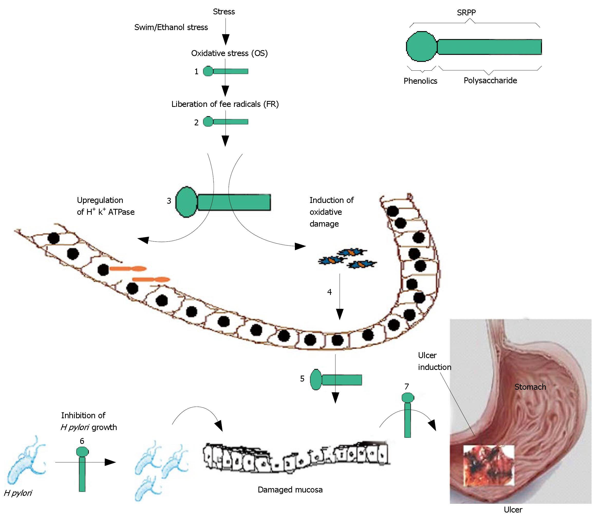
Figure 1 Scheme representing various steps of ulcer pathogenicity and multi-step anti-ulcer action by SRPP (•-); (•) and (-) represents phenolic and polysaccharide portions of SRPP respectively.
Swim/Ethanol stress leading to OS (1) and liberation of FR (2). FR upregulated H+, K+-ATPase (3) and induced oxidative damage to mucosa (4) leading to mucosal damage (5). H pylori may invade on to damaged mucosa and together may cause ulcers (7). SRPP has ability to inhibit steps 1-7 including the growth of H pylori in vitro (6).
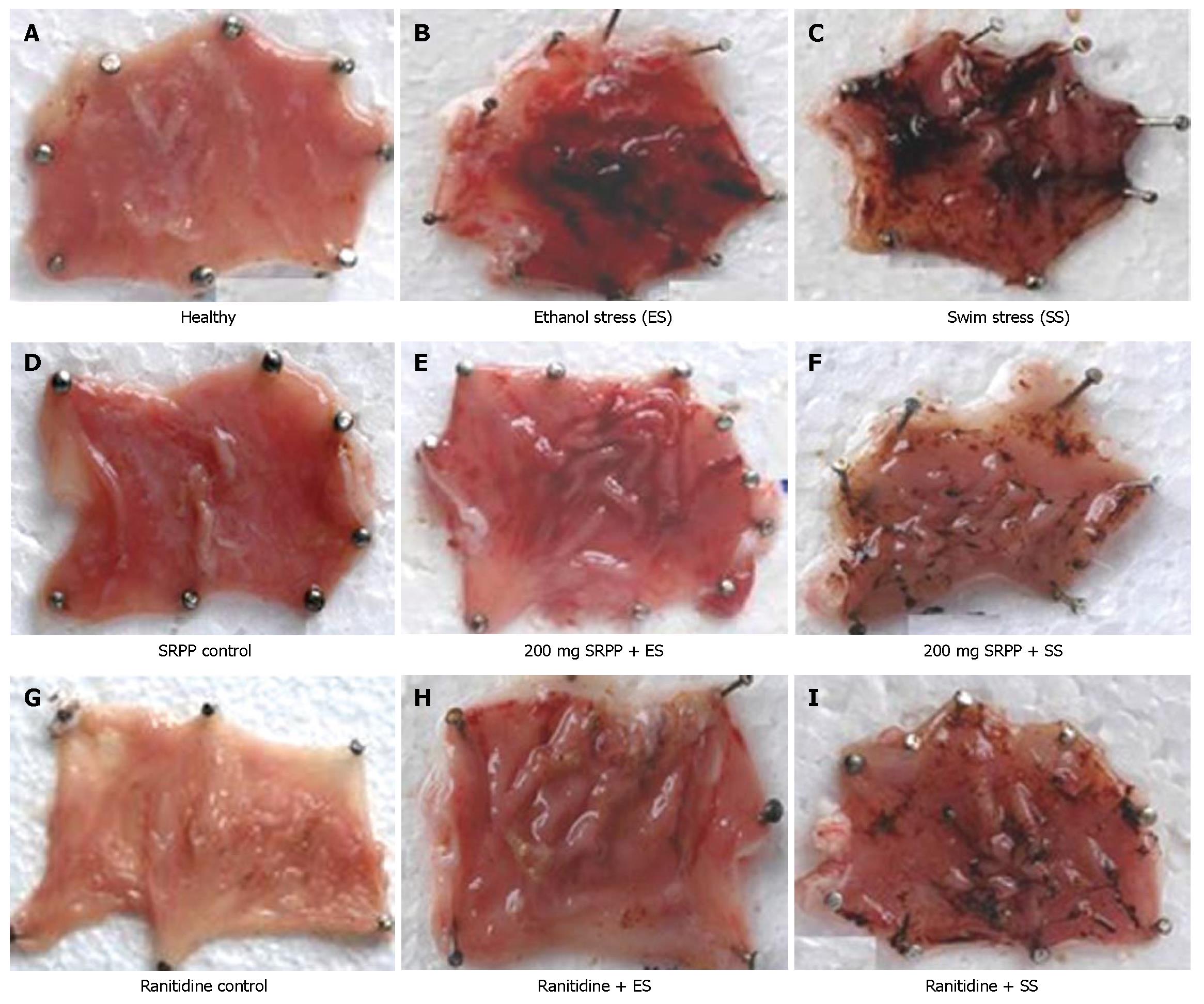
Figure 2 Macroscopic observation of Ulcers in ulcer induced/protected stomachs in swim stress/ethanol stress induced ulcer models; Ulcer was induced in animals by either swim stress (SS) or ethanol stress (ES) in group of pretreated/untreated animals at indicated concentrations.
In healthy (A), SRPP control (D), Ranitidine control (G)-no ulcer lesions or damage in the stomach tissue were observed. In ethanol stress (B) and swim stress (C) induced animals ulcers score were very high. SRPP (E and F) and ranitidine (H and I) treated animals showed reduced stomach lesions.
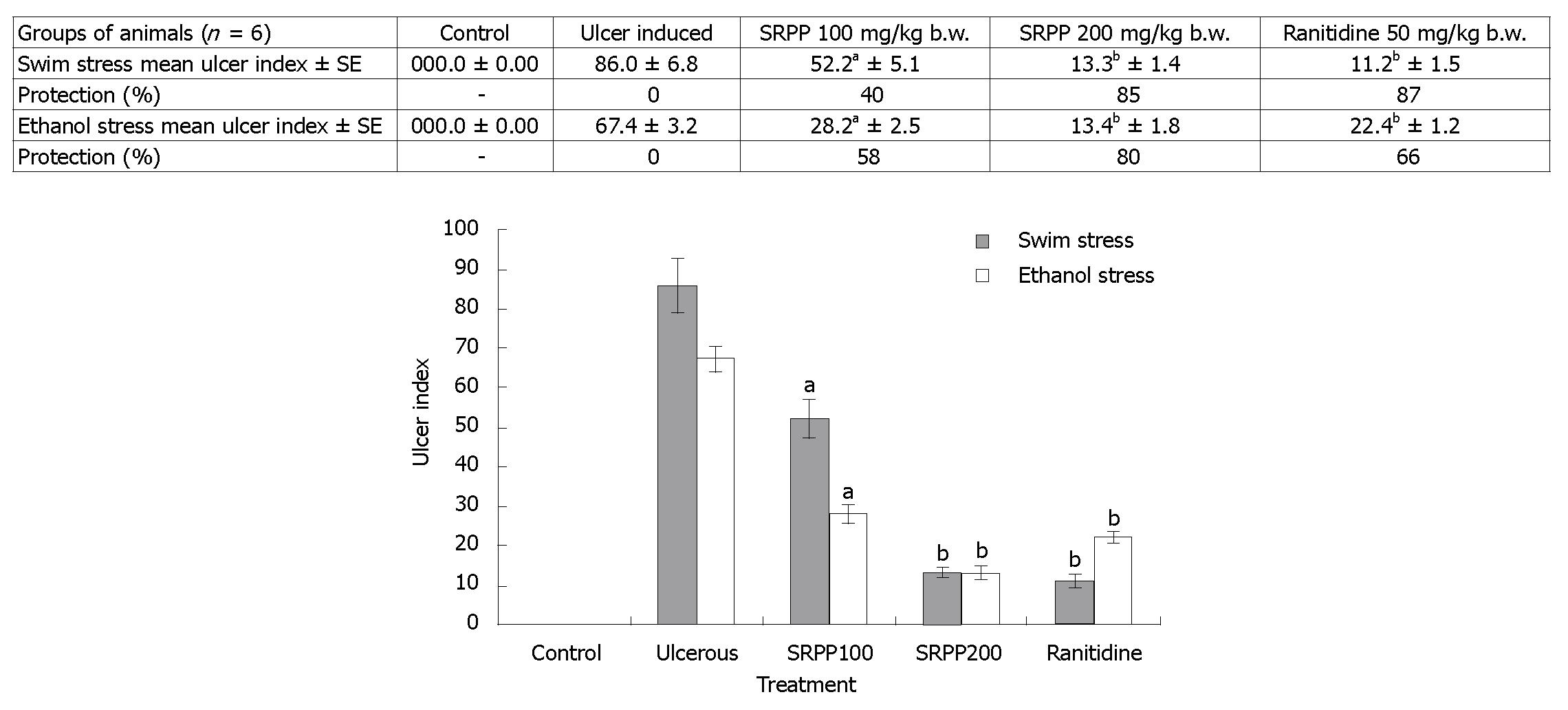
Figure 3 Effect of SRPP on gastric lesions in swim/ethanol stress induced ulcer models; Ulcers were scored as described under the methods and expressed as ulcer index.
Maximum ulcer index observed during stress induction was controlled in a concentration dependent manner. Reduction in ulcer index and percent protection is depicted. aP < 0.05 and bP < 0.01 between ulcerated and treated groups.
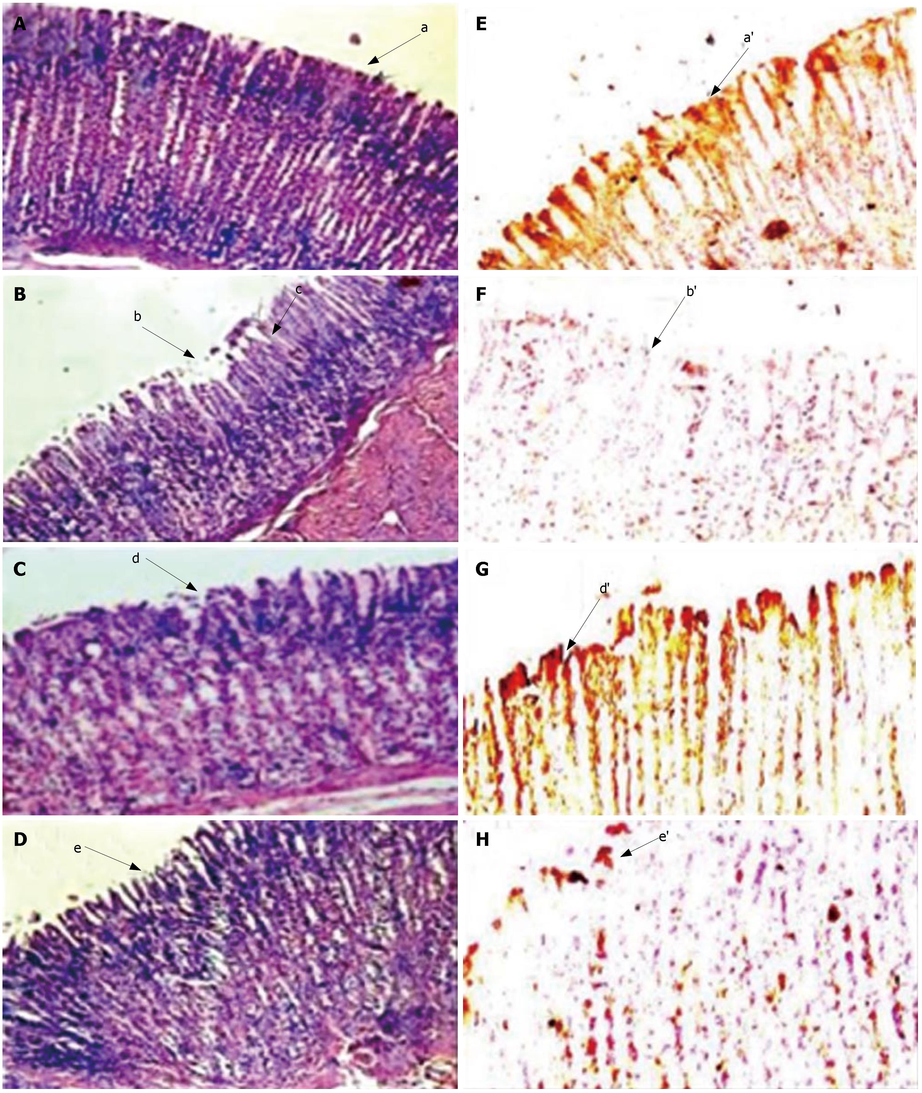
Figure 4 Histopathologic/Immunohistopathologic observation of stomach from ulcer induced/SRPP and Ranitidine treated animals; A-D indicates HE staining sections (× 40), while E-H reveal anti-gastric mucin stained sections (× 40, and magnified the selected portion in computer photoshop).
Control (A, E) shows intact mucosal epithelium with organized glandular structure (a) and intense brown staining for gastric mucin by antibody (a’). Ulcer induction (B, F) showed damaged mucosal epithelium (b) and disrupted glandular structure (c), loss of brown staining (b’) in figure F indicate the loss of gastric mucin. Complete recovery of mucosal damage (d and d’ of C, G) by SRPP and partial recovery by ranitidine (e and e’ of D, H) treatments were observed.
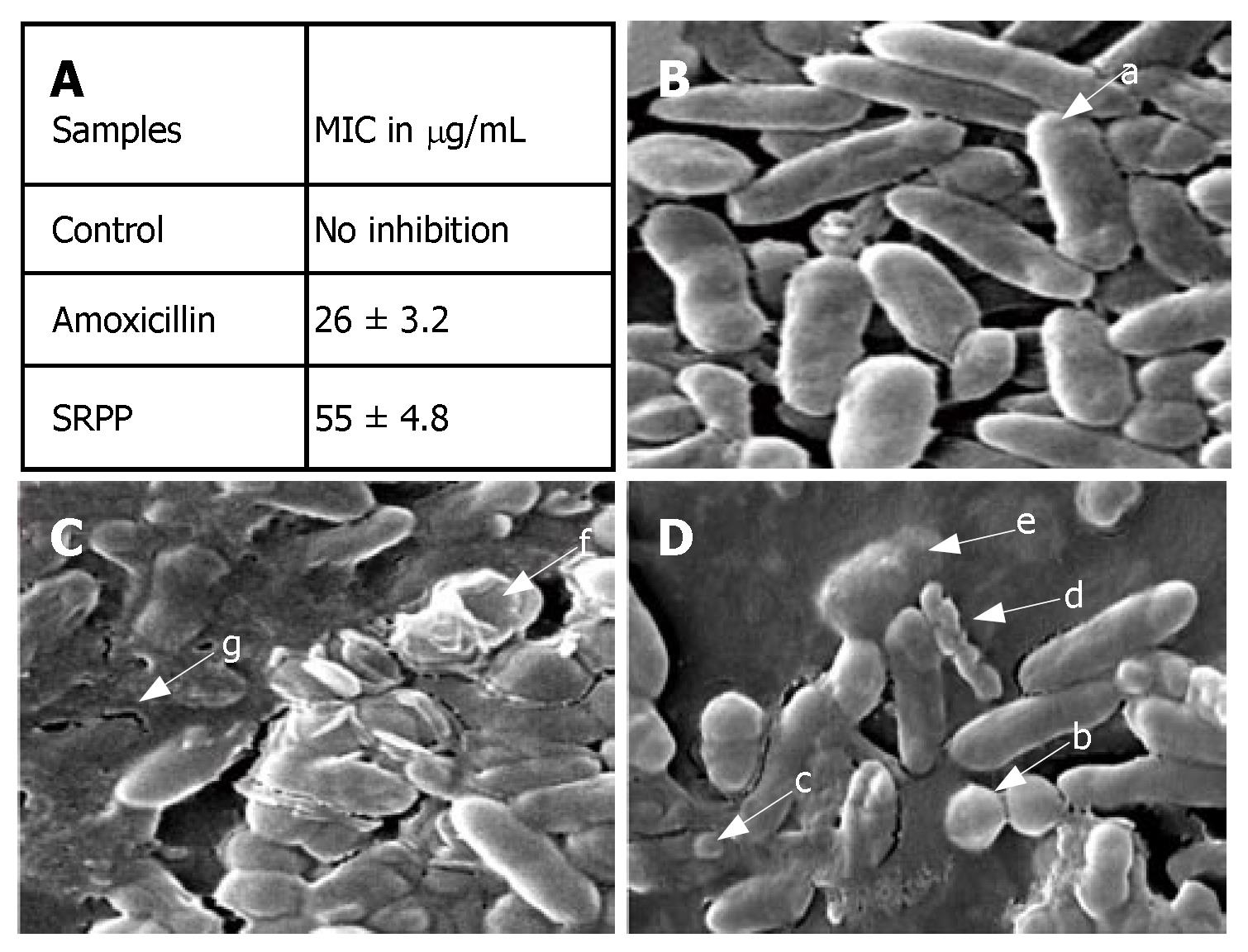
Figure 5 Effect of SRPP on H pylori; Minimum Inhibitory Concentration (MIC) (A) was established by serial dilution technique; B-D indicate the scanning electron microscopic pictures at 15 k magnification of control (B), SRPP (C) and amoxicillin (D) treated H pylori.
Untreated control cultures indicate uniform rod shaped (a) H pylori cells. Amoxicillin treatment showed coccoid form (b), blebbing (c), fragmented (d) and lysed (e) cells. SRPP treatment in addition indicates cavity formation (f) with disrupted structures (g).
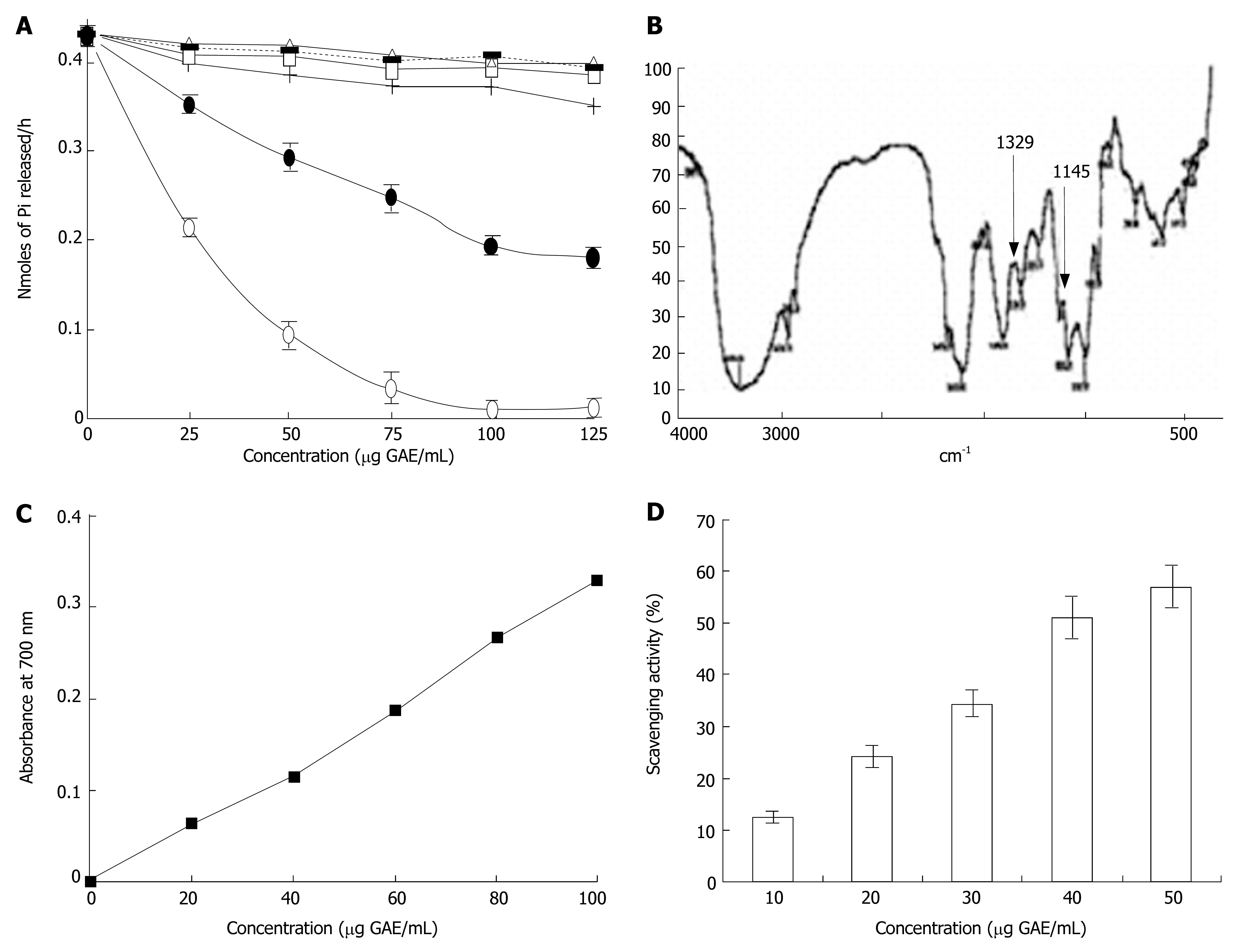
Figure 6 H+, K+-ATPase (A), Fourier Transform Infra-Red Spectroscopy (B), Reducing power (C) and Free radical scavenging activity (D) of SRPP.
A: inhibition of H+, K+-ATPase only by SRPP (•) and not by other polysaccharides of Swallow root; SR water soluble polysaccharide (▪), SR Hemicellulose A (+), Hemicellulose B (□), SR alkali insoluble residue (◊) and inhibition by lansoprazole (o) a known blocker is also depicted in the figure; B: arrow at 1329 and 1145 cm-1 indicate the presence of sulfonamide group in FTIR spectrum. Dose dependent antioxidant activity evaluated as reducing power ability (C) and free radical scavenging ability (D) indicates potential antioxidant activity by phenolics of SRPP.
-
Citation: Srikanta B, Siddaraju M, Dharmesh S. A novel phenol-bound pectic polysaccharide from
Decalepis hamiltonii with multi-step ulcer preventive activity. World J Gastroenterol 2007; 13(39): 5196-5207 - URL: https://www.wjgnet.com/1007-9327/full/v13/i39/5196.htm
- DOI: https://dx.doi.org/10.3748/wjg.v13.i39.5196


