Copyright
©2007 Baishideng Publishing Group Co.
World J Gastroenterol. Sep 7, 2007; 13(33): 4523-4525
Published online Sep 7, 2007. doi: 10.3748/wjg.v13.i33.4523
Published online Sep 7, 2007. doi: 10.3748/wjg.v13.i33.4523
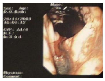
Figure 1 Endoscopic picture showing active bleeding from the lesser curve of the stomach.
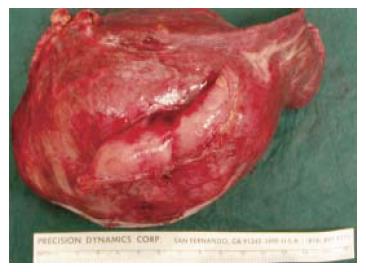
Figure 2 Inferior view of the resected tumor specimen with adherent stomach wall.
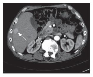
Figure 3 HCC recurrence in the right lobe.
Enhanced axial hepatic arterial-phase CT performed on follow-up after initial resection shows a hypervascular lesion in segment 5/6 suspicious for HCC recurrence (arrow). Incidental note is made of a small cyst in the right kidney.
- Citation: Ong JC, Chow PK, Chan WH, Chung AY, Thng CH, Wong WK. Hepatocellular carcinoma masquerading as a bleeding gastric ulcer: A case report and a review of the surgical management. World J Gastroenterol 2007; 13(33): 4523-4525
- URL: https://www.wjgnet.com/1007-9327/full/v13/i33/4523.htm
- DOI: https://dx.doi.org/10.3748/wjg.v13.i33.4523


