Copyright
©2007 Baishideng Publishing Group Co.
World J Gastroenterol. Sep 7, 2007; 13(33): 4473-4479
Published online Sep 7, 2007. doi: 10.3748/wjg.v13.i33.4473
Published online Sep 7, 2007. doi: 10.3748/wjg.v13.i33.4473
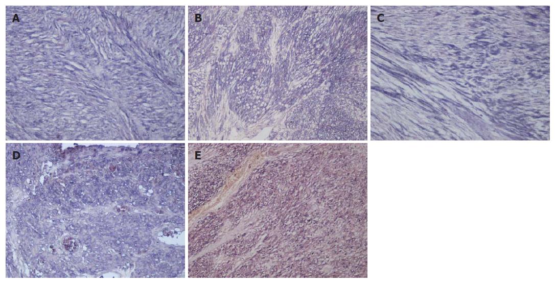
Figure 1 Immunohistochemical staining of Angiopoietin pathway components.
Alkaline phosphatase reaction products demonstrating Ang-1 (A), Ang-2 (B), Ang-4 (C), Tie-1 (D) and Tie-2 (E) expression. Ang-1, -2 and -4 were expressed in the cytoplasm, and Tie-1 and -2 were expressed in both the cytoplasm and the cell membrane of GIST cells (x 200).
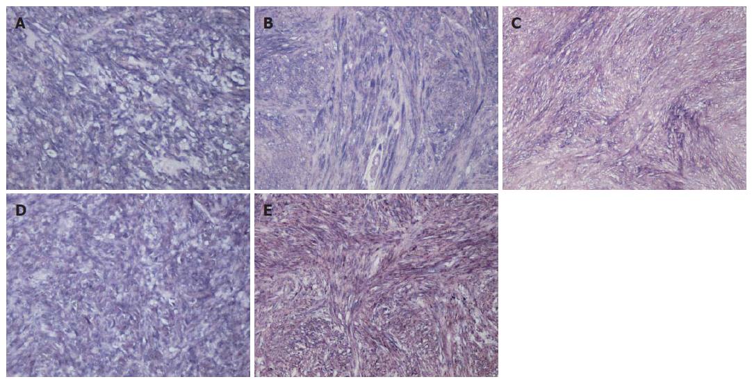
Figure 2 Immunohistochemical staining of human intestinal leiomyomas.
Alkaline phosphatase reaction products demonstrating Ang-1 (A), Ang-2 (B), Ang-4 (C), Tie-1 (D) and Tie-2 (E) expression (x 200).
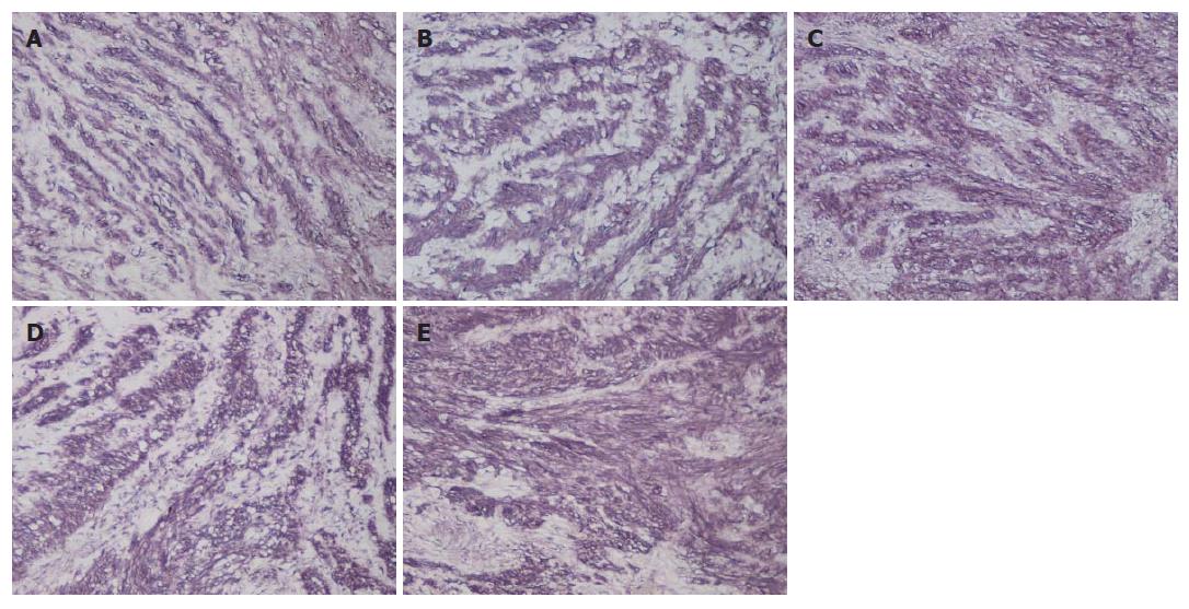
Figure 3 Immunohistochemical staining of human intestinal schwannomas.
Alkaline phosphatase reaction products demonstrating Ang-1 (A), Ang-2 (B), Ang-4 (C), Tie-1 (D) and Tie-2 (E) expression (x 200).
- Citation: Nakayama T, Inaba M, Naito S, Mihara Y, Miura S, Taba M, Yoshizaki A, Wen CY, Sekine I. Expression of Angiopoietin-1, 2 and 4 and Tie-1 and 2 in gastrointestinal stromal tumor, leiomyoma and schwannoma. World J Gastroenterol 2007; 13(33): 4473-4479
- URL: https://www.wjgnet.com/1007-9327/full/v13/i33/4473.htm
- DOI: https://dx.doi.org/10.3748/wjg.v13.i33.4473


