Copyright
©2007 Baishideng Publishing Group Co.
World J Gastroenterol. Aug 14, 2007; 13(30): 4141-4146
Published online Aug 14, 2007. doi: 10.3748/wjg.v13.i30.4141
Published online Aug 14, 2007. doi: 10.3748/wjg.v13.i30.4141
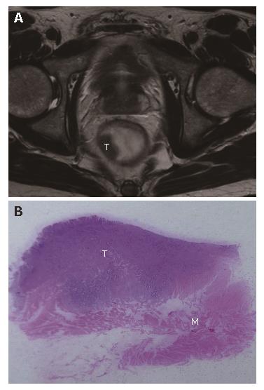
Figure 1 T2-stage rectal cancer in a 58-year-old male patient.
A: Axial T2W-TSE MR image (3500/94); B: Photograph of the corresponding histopathologic slice (hematoxylin-eosin stain; original magnification, x 10), showed T2-stage tumor (T) that was confined within the muscularis propria (M).
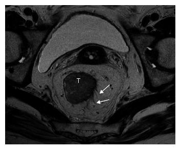
Figure 2 T2-stage rectal cancer overstaged at MR imaging (3000/98) as a T3-stage tumor in a 70-year-old male patient.
MR image depicted tumor (T) with speculations (white arrow) which turned out to desmoplastic reaction without tumor cells at histology.
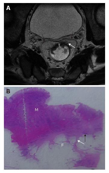
Figure 3 A: T3-stage rectal cancer without mesorectal fascia involvement in a 64-year-old female patient.
Axial T2W-TSE MR image (3000/98) showed tumor in anterior rectal wall (T) with transmural spiculation (black arrow) from tumor into perirectal fat, and the distance to mesorectal fascia (white arrow) is measured ≤2 mm; B: T3-stage rectal cancer with involved mesorectal fascia in a 79-year-old male patient. Axial T2W-TSE MR image (3000/98) showed tumor (T) extending to mesorectal fascia (white arrows); C: T3-stage rectal cancer with involved mesorectal fascia in a 65-year-old male patient. Axial T2W-TSE MR image (3000/98) showed heterogeneous strands are noted in the perirectal fat tissues with the thicken mesorectal fascia; D: T3-stage rectal cancer with extramural deposits in a 62-year-old female patient. Axial T2W-TSE MR image (3000/98) showed extramural deposits (T) in the perirectal space with irregular shape.
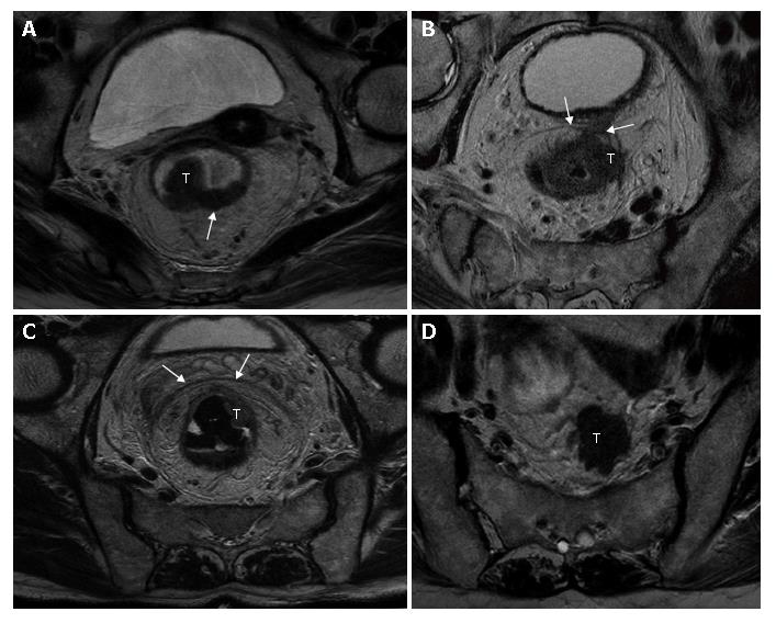
Figure 4 A: T3-stage rectal cancer without mesorectal fascia involvement in a 57-year-old female patient.
Axial T2W-TSE MR image (3000/98) manifested bulky tumor with broad-based nodular (T) with clear margin (white arrows); B: T3-stage rectal cancer with involved mesorectal fascia in a 79-year-old male patient. Axial T2W-TSE MR image (3000/98) showed tumor (T) extending to mesorectal fascia (white arrows); C: T3-stage rectal cancer with involved mesorectal fascia in a 65-year-old male patient. Axial T2W-TSE MR image (3000/98) showed heterogeneous strands are noted in the perirectal fat tissues with the thicken mesorectal fascia; D: T3-stage rectal cancer with extramural deposits in a 62-year-old female patient. Axial T2W-TSE MR image (3000/98) showed extramural deposits (T) in the perirectal space with irregular shape.
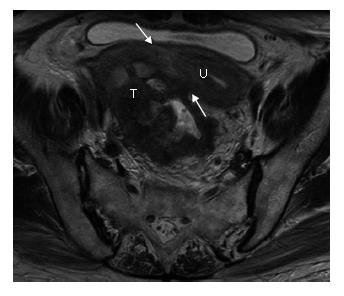
Figure 5 T4-stage rectal cancer with fixation to uterus in a 76-year-old female patient.
Axial T2W-TSE MR image (3000/98) showed direct invasion (white arrows) of tumour (T) into uterus (U).
- Citation: Rao SX, Zeng MS, Xu JM, Qin XY, Chen CZ, Li RC, Hou YY. Assessment of T staging and mesorectal fascia status using high-resolution MRI in rectal cancer with rectal distention. World J Gastroenterol 2007; 13(30): 4141-4146
- URL: https://www.wjgnet.com/1007-9327/full/v13/i30/4141.htm
- DOI: https://dx.doi.org/10.3748/wjg.v13.i30.4141


