Copyright
©2007 Baishideng Publishing Group Co.
World J Gastroenterol. Aug 14, 2007; 13(30): 4080-4084
Published online Aug 14, 2007. doi: 10.3748/wjg.v13.i30.4080
Published online Aug 14, 2007. doi: 10.3748/wjg.v13.i30.4080
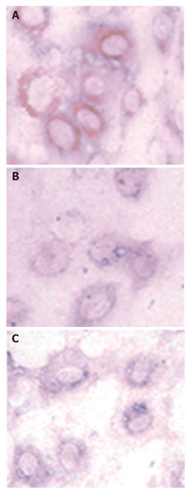
Figure 1 HCV NS5A protein staining of Huh7 cells after 48 h transfection (DAB, × 400).
A: PCNS5A-transfected cells; B: PRC/CMV-transfected cells; C: Non-transfected cells.
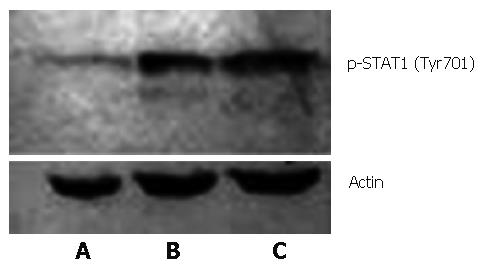
Figure 2 Tyr701 phosphorylated STAT1 in Huh7 cells induced by IFNα-2b (10 000 U/mL) (Western blot).
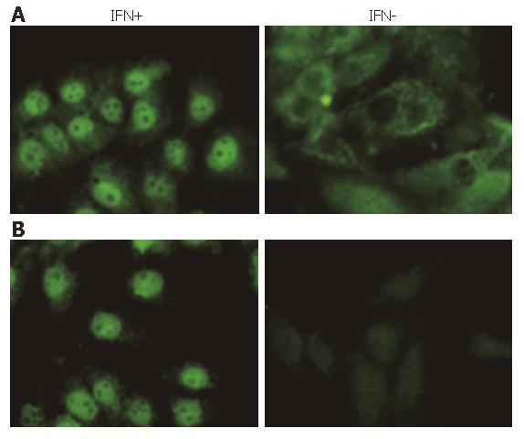
Figure 3 STAT1 or Phosphorylated STAT1 (Tyr701) in Huh7 cells stimulated by IFNα-2b (10 000 U/mL) for 30 min (FITC, × 400).
A: STAT1; B: Tyr701.
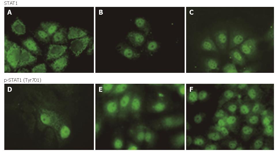
Figure 4 STAT1 or phosphorylated STAT1 in cytoplasm and nuclei of Huh7 cells after 30 min IFN induced (FITC, × 400): A and D: PCNS5A-transfected; B and E: PRC/CMV-transfected; C and F: non-transfected.
Figure 5 Phosphorylated STAT1 from IFN induced Huh7 cells (FITC, × 400): A: PCNS5A-transfected; B: PRC/CMV-transfected; C: Non-transfected.
- Citation: Gong GZ, Cao J, Jiang YF, Zhou Y, Liu B. Hepatitis C Virus non-structural 5A abrogates signal transducer and activator of transcription-1 nucleartranslocation induced by IFN-α through dephosphorylation. World J Gastroenterol 2007; 13(30): 4080-4084
- URL: https://www.wjgnet.com/1007-9327/full/v13/i30/4080.htm
- DOI: https://dx.doi.org/10.3748/wjg.v13.i30.4080


