Copyright
©2007 Baishideng Publishing Group Co.
World J Gastroenterol. Jan 21, 2007; 13(3): 483-485
Published online Jan 21, 2007. doi: 10.3748/wjg.v13.i3.483
Published online Jan 21, 2007. doi: 10.3748/wjg.v13.i3.483
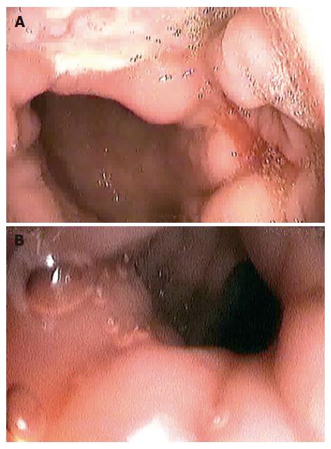
Figure 1 Upper endoscopy showing large and deep ulceration in the posterior gastric wall (A) and ulceration with perforation in the small intestine (B).
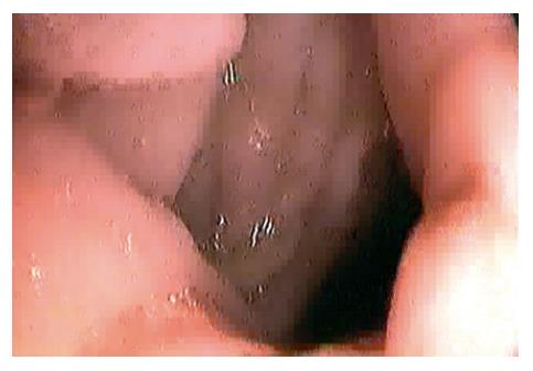
Figure 2 Small intestine loop reached by endoscope through spontaneously eloped gastrojejunal fistula.
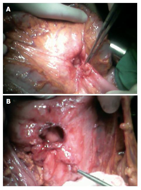
Figure 3 Hole in mescaline with gastrojejunal fistula (A) and defect in posterior gastric wall after resection of fistula and deep gastric ulcer (B).
- Citation: Ćulafić ĐM, Matejić OD, Đukić VS, Vukčević MD, Kerkez MD. Spontaneous gastrojejunal fistula is a complication of gastric ulcer. World J Gastroenterol 2007; 13(3): 483-485
- URL: https://www.wjgnet.com/1007-9327/full/v13/i3/483.htm
- DOI: https://dx.doi.org/10.3748/wjg.v13.i3.483


