Copyright
©2007 Baishideng Publishing Group Co.
World J Gastroenterol. Jan 21, 2007; 13(3): 474-477
Published online Jan 21, 2007. doi: 10.3748/wjg.v13.i3.474
Published online Jan 21, 2007. doi: 10.3748/wjg.v13.i3.474
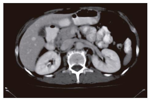
Figure 1 Abdominal computed tomograph revealed a heterogeneously enhanced, multicystic structure, 3 cm in diameter, cephalad to the pancreatic head.
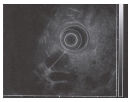
Figure 2 EUS with a radial echoendoscope revealed a large, 2-2.
5 cm anoechoic, homogenous, well-defined mass which lay adjacent to the neck of the pancreas.
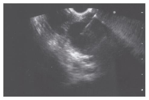
Figure 3 EUS-guided FNA of the peripancreatic lesion was performed using a linear-array echoendoscope.
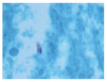
Figure 4 Staining with Ziel Nielson within 30 min yielded a positive result for acid-fast bacilli.
- Citation: Boujaoude JD, Honein K, Yaghi C, Ghora C, Abadjian G, Sayegh R. Diagnosis by endoscopic ultrasound guided fine needle aspiration of tuberculous lymphadenitis involving the peripancreatic lymph nodes: A case report. World J Gastroenterol 2007; 13(3): 474-477
- URL: https://www.wjgnet.com/1007-9327/full/v13/i3/474.htm
- DOI: https://dx.doi.org/10.3748/wjg.v13.i3.474


